Main menu
Common skin conditions

NEWS
Join DermNet PRO
Read more
Quick links

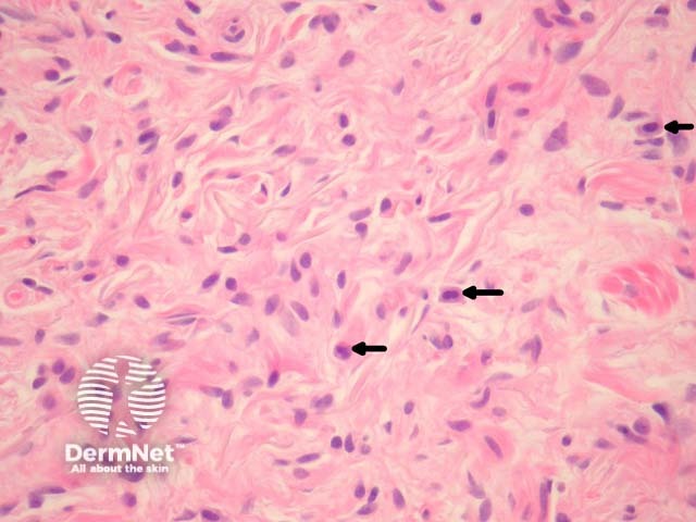
Figure 4
Keywords: Neurofibroma, Histopathology-image, Pathology
In a neurofibroma, sections show a non-encapsulated lesion in the dermis (figure 1). These lesions represent a proliferation of all elements of peripheral nerves. The cells have wavy serpentine nuclei and pointed ends (figures 2-4). There is stromal mucin deposition and fibroplasia (figures 2-4). Mast cells are usually easy to find (figure 4, arrow). Axons course through the tumour and though difficult to see on routine sections may be illustrated with immunohistochemical stains for neurofilament.
© DermNet
You can use or share this image if you comply with our image licence. Please provide a link back to this page.
For a high resolution, unwatermarked copy contact us here. Fees apply.
Source: dermnetnz.org