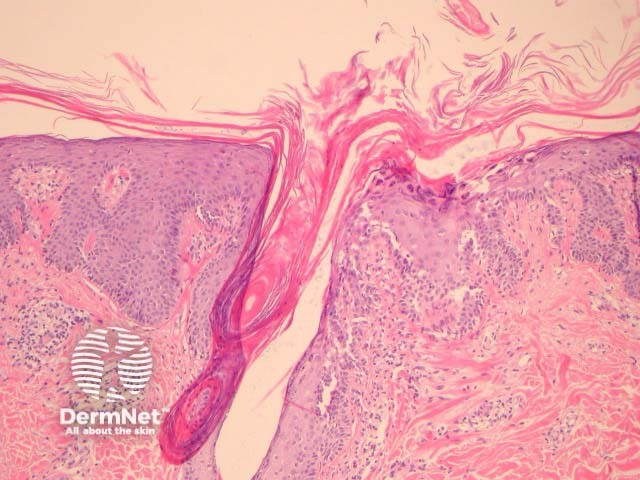
Figure 3
Keywords: Histopathology-image
In PRP, the epidermis shows regular acanthosis and psoriasiform hyperplasia (figures 1-3). The horn is thickened with parakeratotic foci between orthokeratosis both vertically and horizontally (not seen clearly in the presented images). The hyperkeratosis tracks down the openings of follicular structures forming follicular plugs (figures 1-3). The dermis may contain a mixed sparse infiltrate which may be lichenoid. Rarely, foci of acantholysis may be seen (figure 3) and some authors have made associations with Darier disease.
© DermNet
You can use or share this image if you comply with our image licence. Please provide a link back to this page.
For a high resolution, unwatermarked copy contact us here. Fees apply.
Source: dermnetnz.org

