Figure 2
Keywords: Urachal cyst, Histopathology-image, Pathology
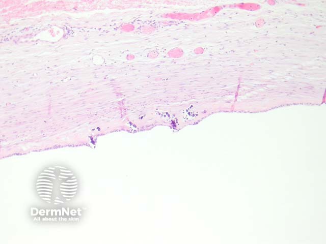
These are lined by urothelium, cuboidal, flat or atrophic epithelia (figures 1, 2, 3). The cyst may be located at any point between the bladder and the umbilicus. The surrounding dermis or soft tissue often shows fibrosis, calcification and reactive changes (figures 1, 2, 3)
© DermNet
You can use or share this image if you comply with our image licence. Please provide a link back to this page.
For a high resolution, unwatermarked copy contact us here. Fees apply.
Source: dermnetnz.org

