Figure 4
Keywords: Pleomorphic lipoma, Histopathology-image, Pathology
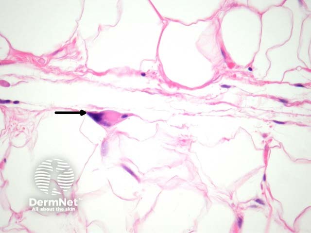
Sections of pleomorphic lipoma show a circumscribed fatty tumour with mature fat admixed with more cellular areas (figure 1). There are often mucinous and spindled areas similar to those seen in spindle cell lipoma. There are intermixed lipoblast-like cells which are giant, may be multinucleated, and often referred to as floret giant cells (figures 2-4, arrows). The nuclei of the giant cells are smudged may be places around the periphery of the cell and have smudged nuclei (otherwise called “floret giant cells”). The cytoplasm is solid and eosinophilic (figures 2-4, arrows).
© DermNet
You can use or share this image if you comply with our image licence. Please provide a link back to this page.
For a high resolution, unwatermarked copy contact us here. Fees apply.
Source: dermnetnz.org

