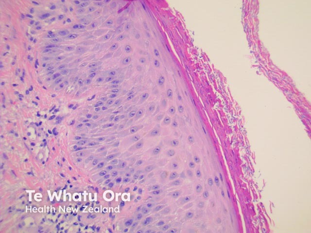
Figure 2
Keywords: Tinea incognita, Histopathology-image, Pathology
In tinea incognito, the epidermis is often mildly spongiotic, and the dermal inflammatory infiltrate is less heavy than usual dermatophytosis. Multiple branched, septate hyphae and small spores are present in the stratum corneum. These can often be seen on H-E stain (figures 1, 2, 3) but special stains are often needed to reveal the fungi and demonstrate their morphology.
© DermNet
You can use or share this image if you comply with our image licence. Please provide a link back to this page.
For a high resolution, unwatermarked copy contact us here. Fees apply.
Source: dermnetnz.org

