Figure 3
Keywords: Histopathology-image
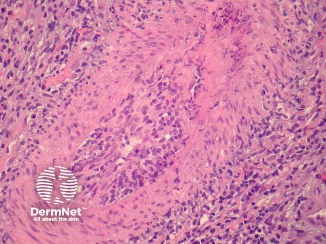
Histological features of systemic and localised cutaneous forms of polyarteritis nodosa are similar. A deep biopsy is preferred as cutaneous polyarteritis nodosa involves medium-sized vessels in the deep dermis and subcutis (Figure 1). Early lesions show fibrinoid necrosis with thickening and infiltration of the vessel wall. Neutrophils, eosinophils and lymphocytes are present (Figures 2,3). Leucocytoclasis may be present. Thrombi and aneurysmal change may occur and lead to necrosis of the overlying epithelium. In mature lesions vessel occlusion occurs secondary to intimal and mural fibrosis. Lesions at various stages are characteristic and changes are discontinuous with uninvolved skip lesions between affected segments.
© DermNet
You can use or share this image if you comply with our image licence. Please provide a link back to this page.
For a high resolution, unwatermarked copy contact us here. Fees apply.
Source: dermnetnz.org

