Go to dermatofibroma topic page
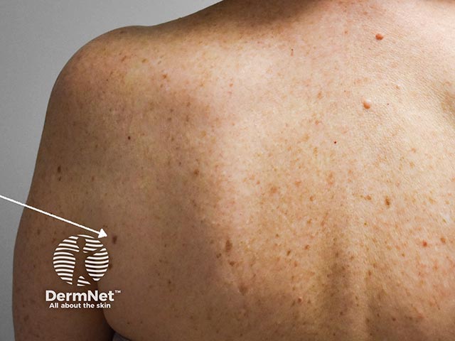
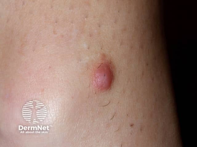
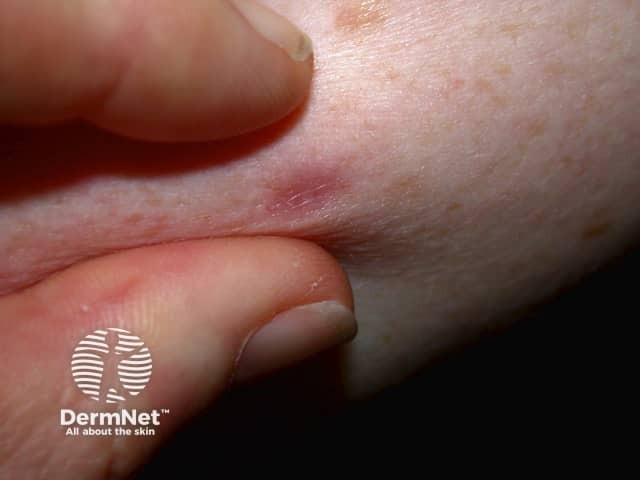
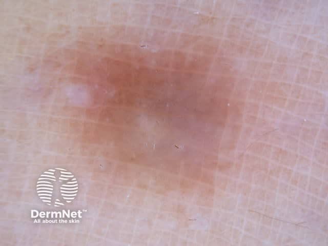
A dermatofibroma on the shoulder - they are often hyperpigmented and 'dip' into the skin. This can be accentuated by pinching (the pinch sign) (DF-patient1)
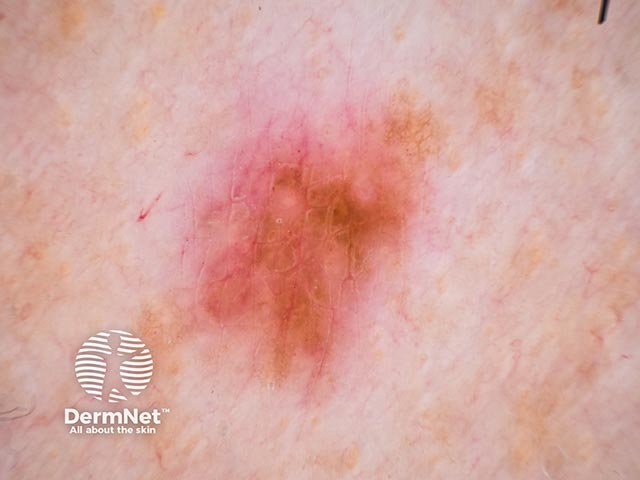
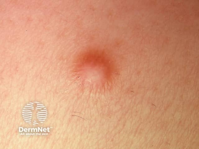
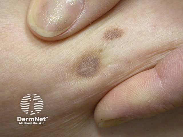
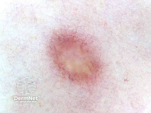
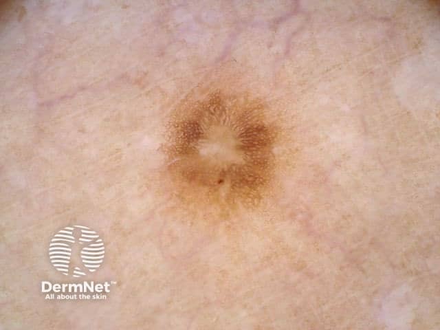
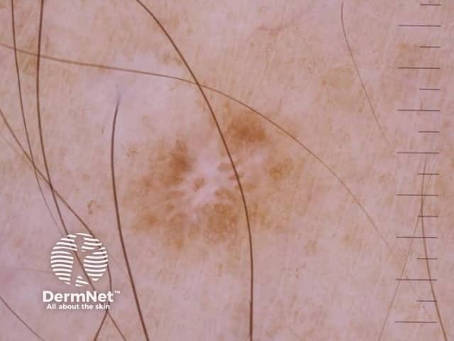
Dermoscopy of a dermatofibroma - there is often central pallor and overlying lentiginous hyperplasia (DF-patient1)
Dermatofibroma macro images
Dermatofibroma dermoscopy images
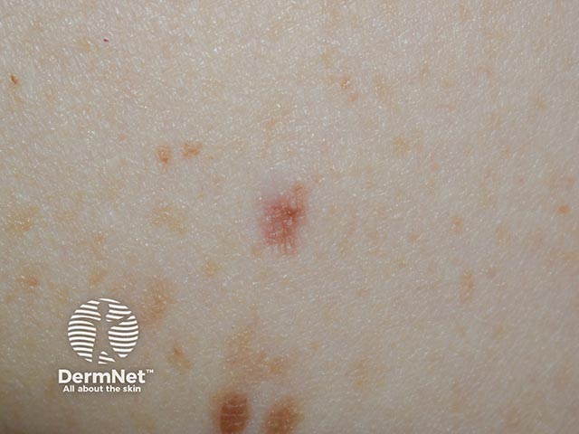
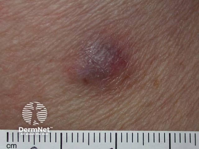
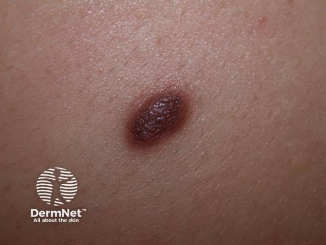
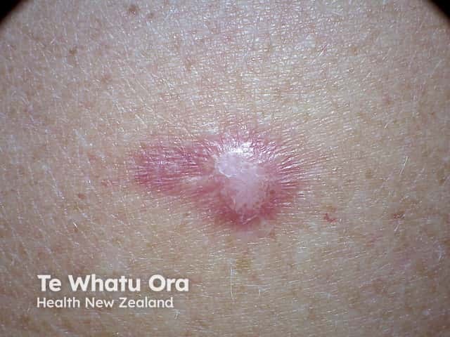
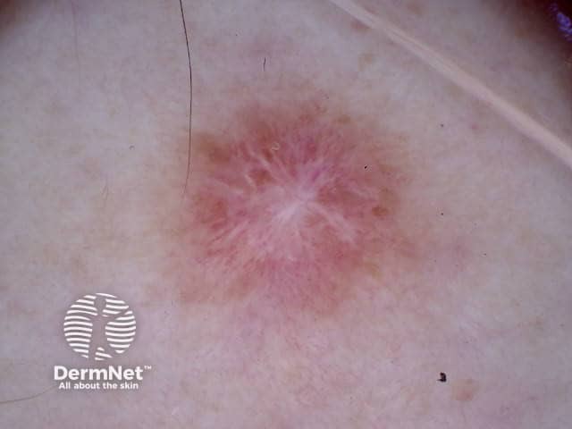
Dermatoscopy of dermatofibroma
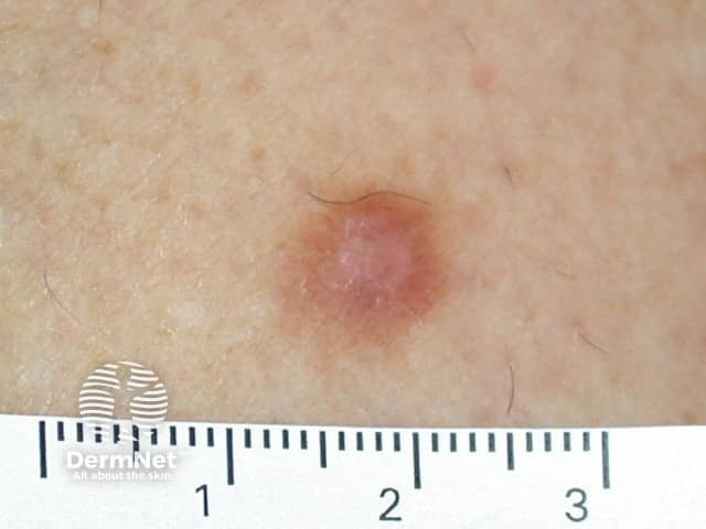
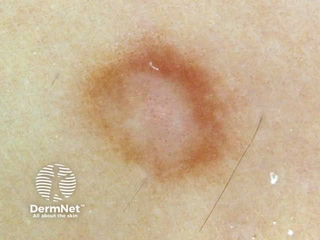
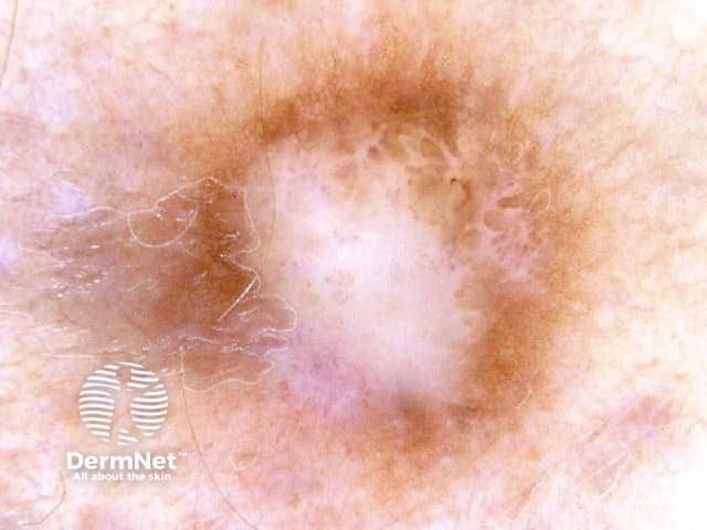
Dermoscopy. Peripheral network, central white area: dermatofibroma
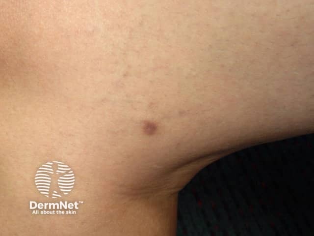
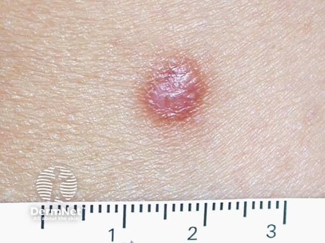
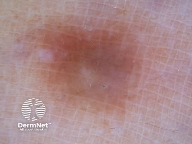
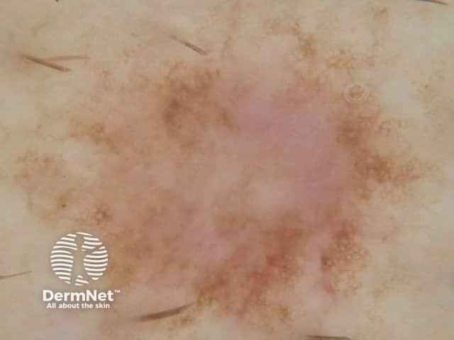
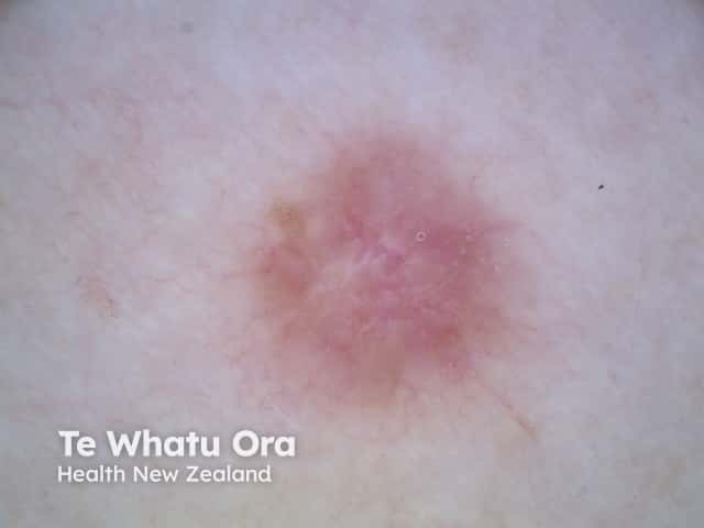
Dermoscopy. Bland, structureless pigmentation: dermatofibroma
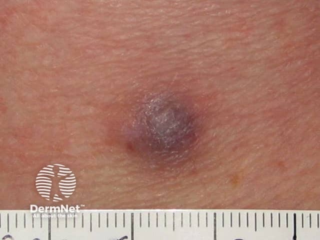
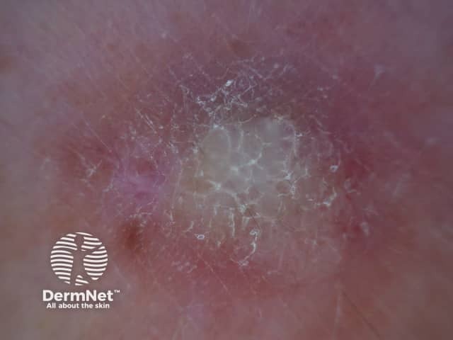
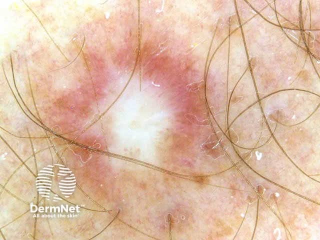
Dermoscopy. Prominent central white area: dermatofibroma
Go to dermatofibroma topic page