Go to the superficial spreading melanoma topic page
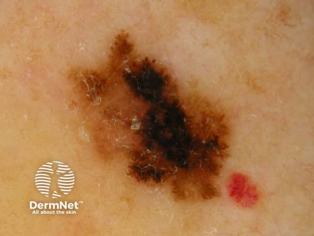
A thin superficial spreading malignant melanoma - there is irregular variability of the pigmentation
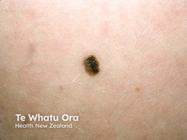
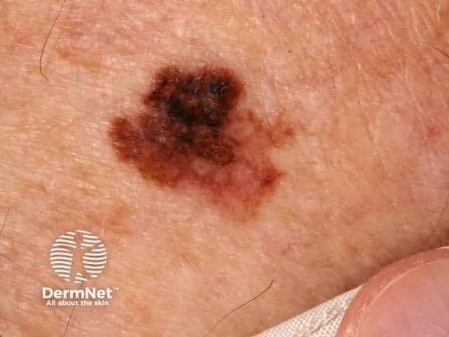
Superficial spreading malignant melanoma - an ugly duckling with an irregular edge and pigmentation (SSM-patient1)
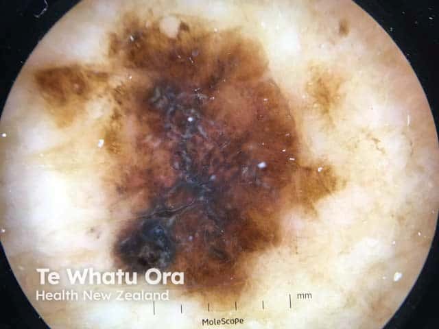
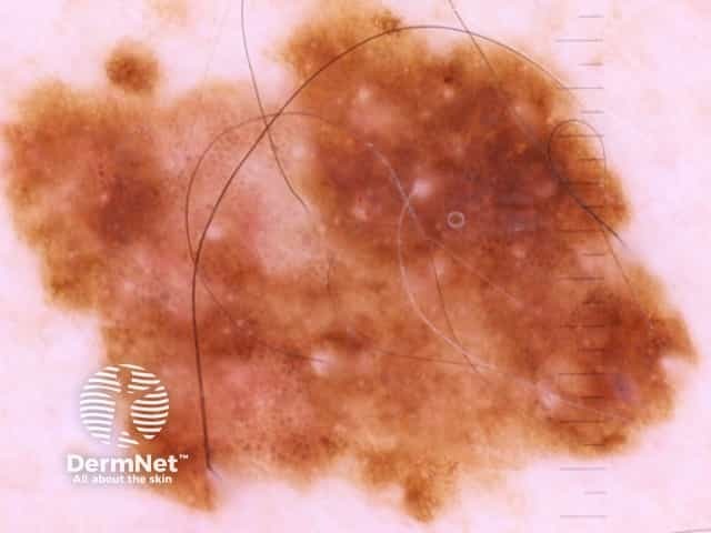
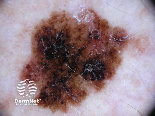
Dermoscopy of a superficial spreading malignant melanoma - irregular edge and pigmentation and a blue-grey veil (SSM-patient1)
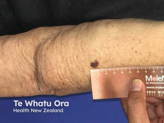
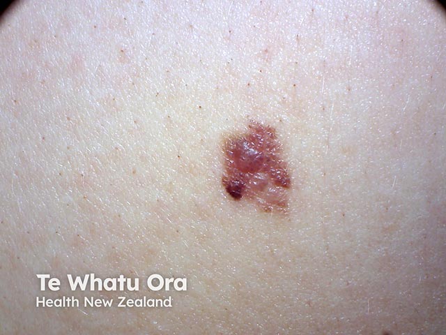
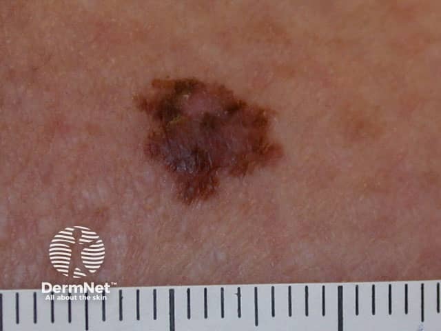
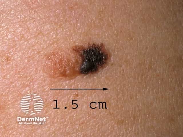
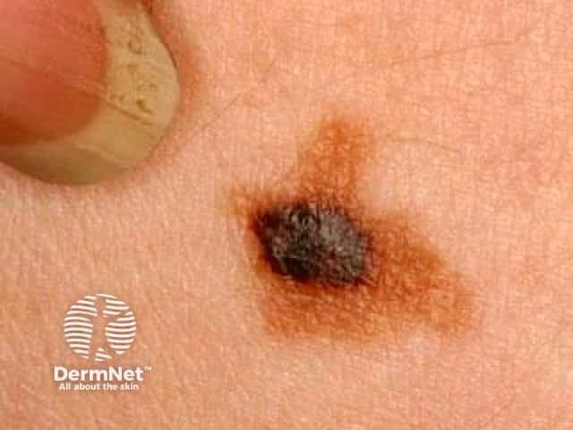
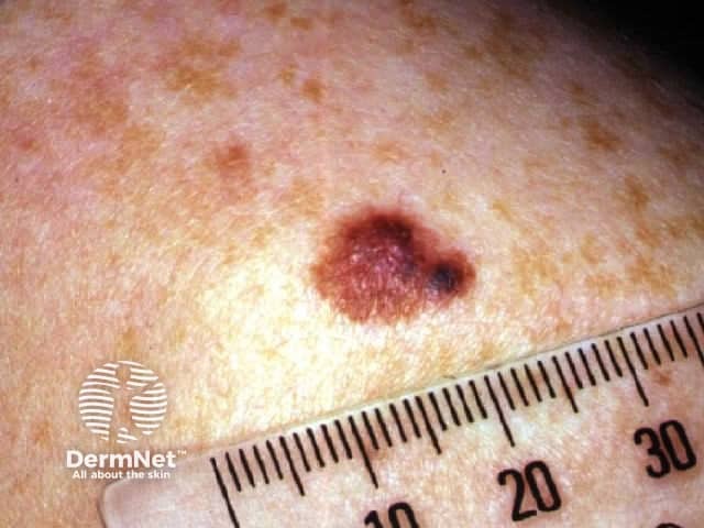
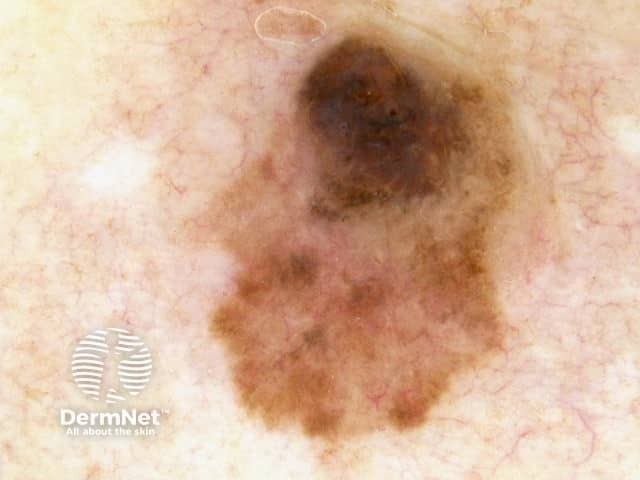
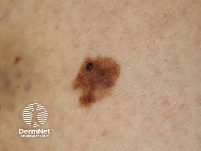
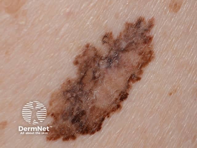
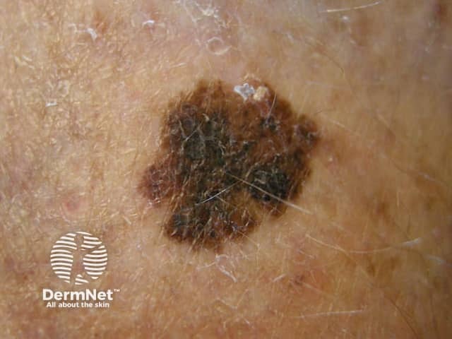
An irregularly marginated and pigmented ugly duckling - an 0.8 mm Breslow thickness superficial spreading malignant melanoma
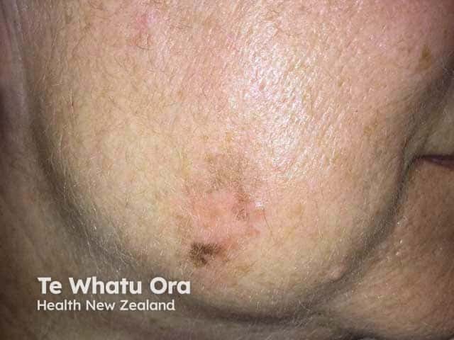
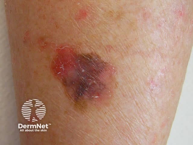
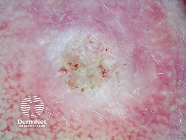
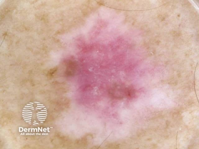
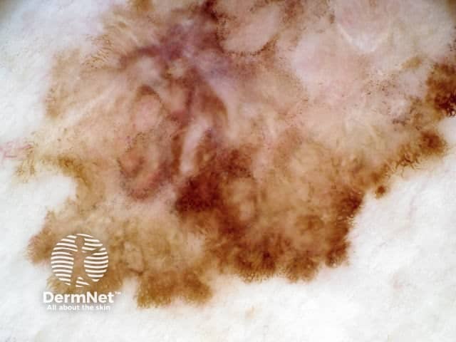
An irregular mole with variable colour and a white area representing regression of a superficial spreading malignant melanoma
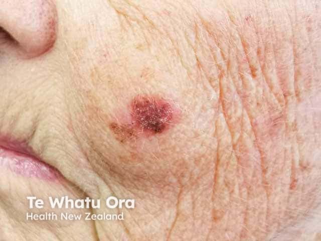
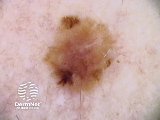
An irregularly marginated and pigmented lesion on the cheek - a superficial spreading malignant melanoma
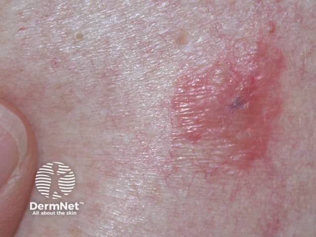
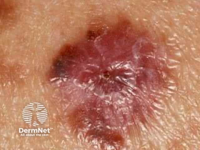
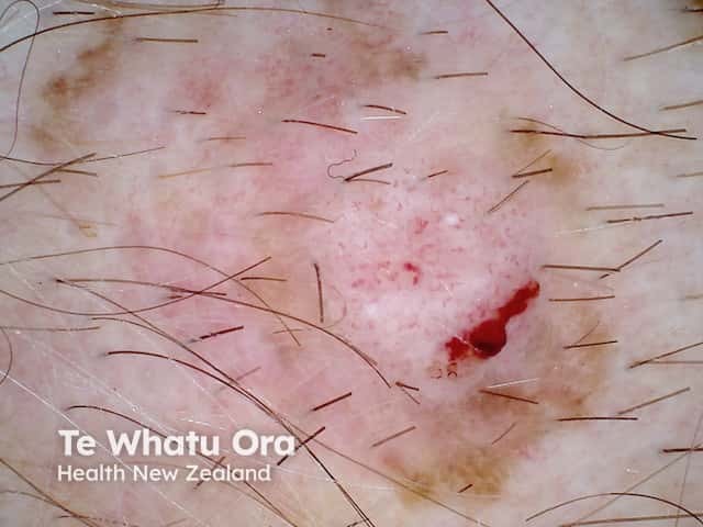
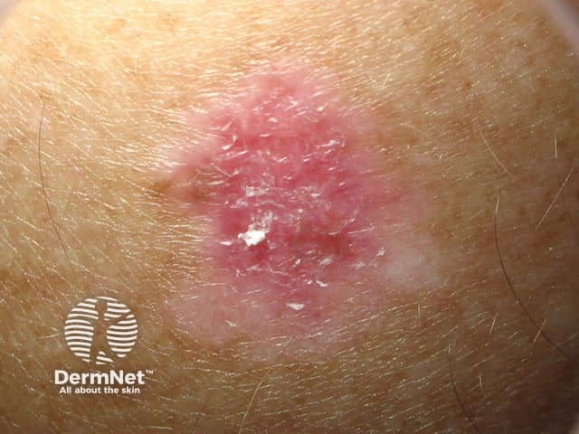
A melanoma on the cheek showing clinical evidence of central regression
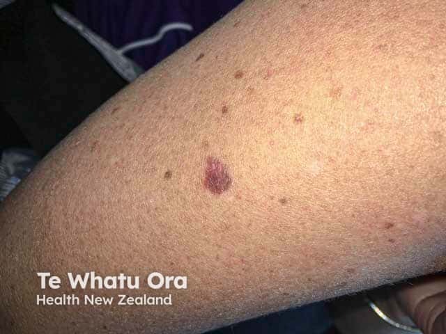
An ugly duckling pigmented lesion on the arm - a superficial spreading malignant melanoma
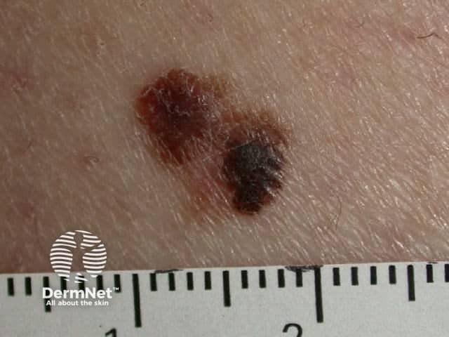
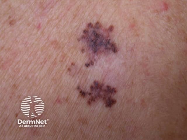
Multiple areas of notching of the border of this ugly duckling lesion - a malignant melanoma (SSM-patient2)
Multiple areas of notching of the border of this ugly duckling lesion - a malignant melanoma (SSM-patient2)
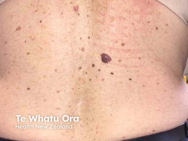
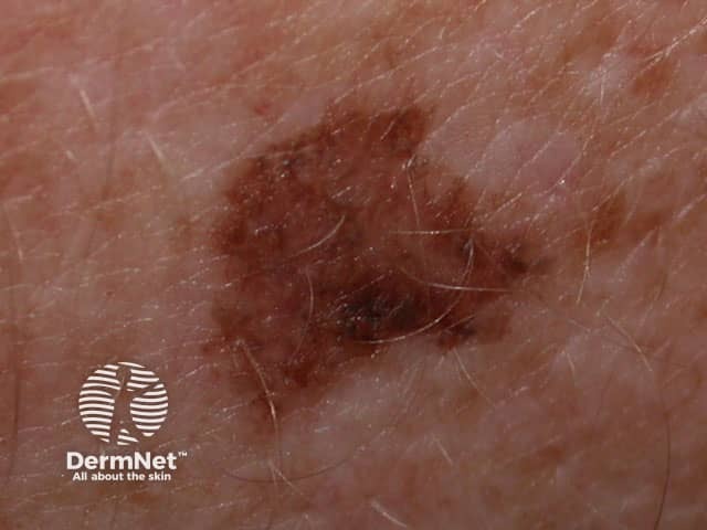
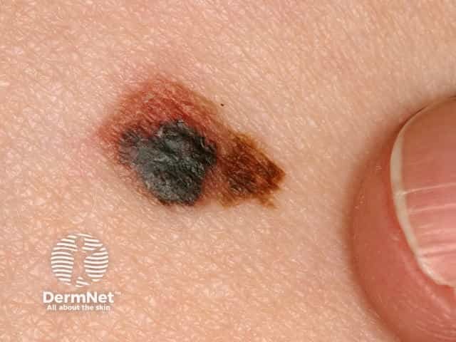
Irregular edge, asymmetry and irregular pigmentation of a melanoma on the back
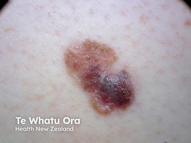
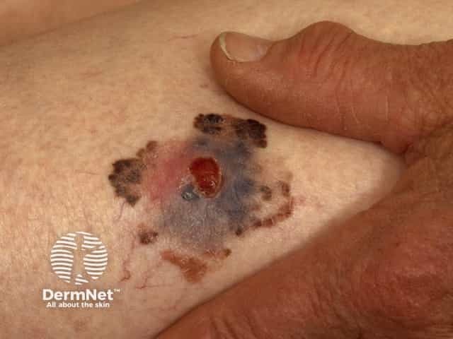
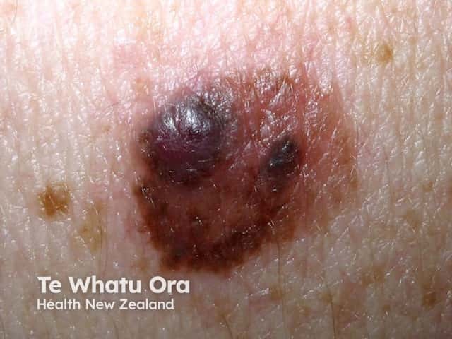
A nodular melanoma arising within a superficial spreading melanoma on the back (SSM-patient3)
An amelanotic nodular melanoma arising within a superficial spreading melanoma on the back (SSM-patient3)
An amelanotic nodular melanoma arising within a superficial spreading melanoma on the back (SSM-patient3)
An amelanotic nodular melanoma arising within a superficial spreading melanoma on the back (SSM-patient3)
An amelanotic nodular melanoma arising within a superficial spreading melanoma on the back (SSM-patient3)
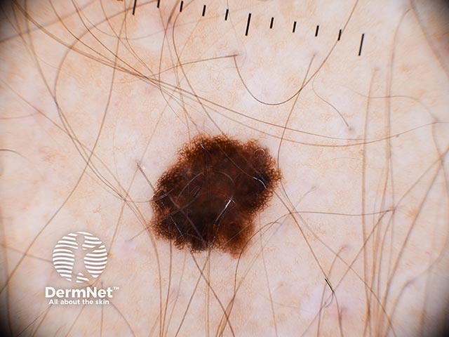
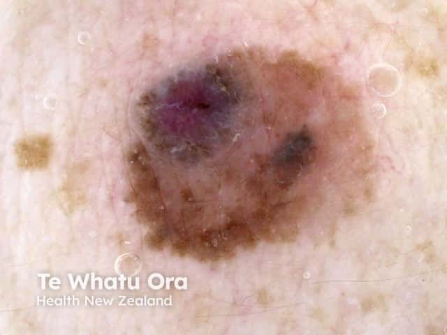
Dermoscopic image of a superficial spreading melanoma on the forearm (SSM-patient4)
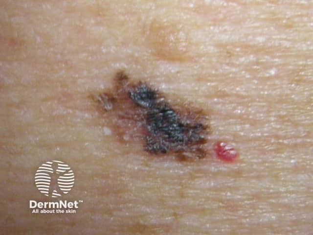
A melanoma in situ on the lower back - dark brown and black in colour with irregularity and notching of the border (SSM-patient5)
A melanoma in situ on the lower back - dark brown and black in colour with irregularity and notching of the border, and an atypical pigment network (SSM-patient5)
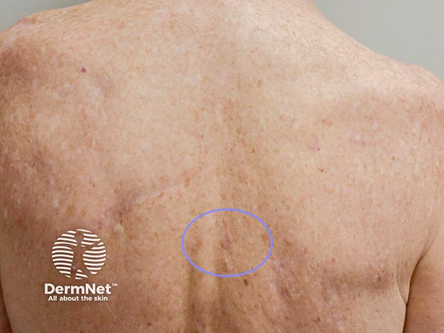
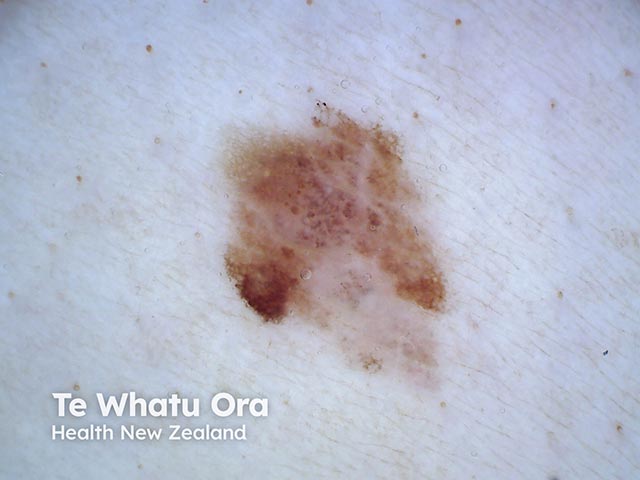
A hypomelanotic macule on the mid back - excision showed a thin superficial spreading melanoma (SSM-patient6)
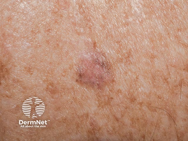
Close-up of a hypomelanotic thin superficial spreading melanoma on the back (SSM-patient6)
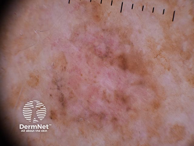
Dermoscopy of a hypomelanotic superficial spreading melanoma - irregular edge, atypical vessels and irregular grey dots (SSM-patient6)
A changing irregularly-pigmented lesion on the ankle - an in situ melanoma (SSM-patient7)
An in situ melanoma showing irregular pigmentation and border, and peripheral structureless areas (SSM-patient7)
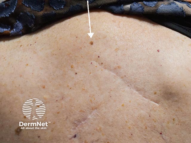
A superficial spreading melanoma on the back in a patient with a personal history of melanoma (SSM-patient8)
A superficial spreading melanoma on the back in a patient with a personal history of melanoma (SSM-patient8)
A superficial spreading melanoma on the back showing irregular grey dots (SSM-patient8)
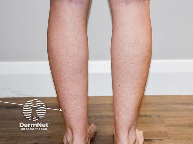
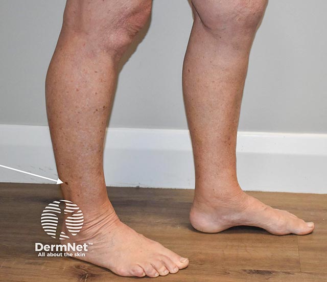
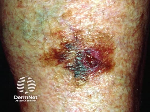
An in situ melanoma on the right lateral calf with early invasion (SSM-patient9)
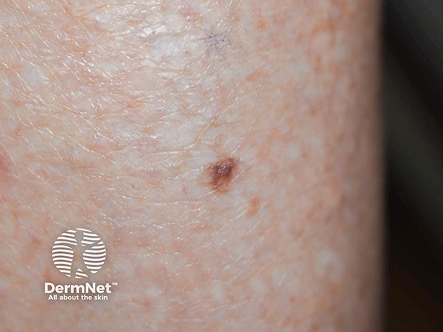
A thin invasive melanoma arising within an in situ lesion on the calf - note the irregular edge and notch (SSM-patient9)
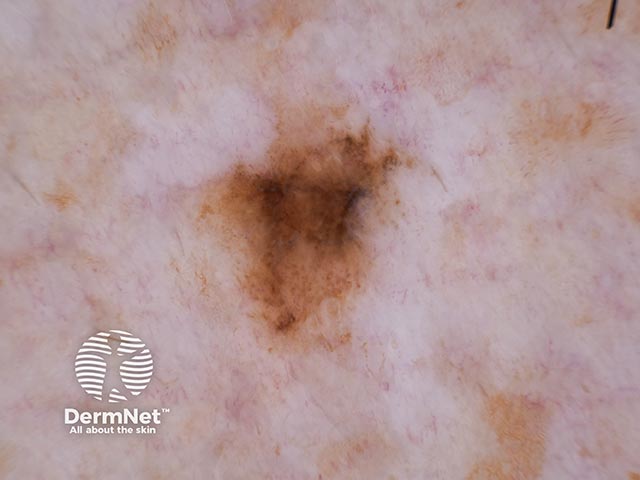
A thin invasive melanoma - note the chaotic arrangement and irregular pigment net with blotches and grey-white area at 3 o'clock (SSM-patient9)
A changing melanocytic lesion - linear vessels are seen centrally (SSM-patient13)
Melanoma in situ - a changing pigmented melanocytic lesion on the back (SSM-patient13)
Dermoscopy of a nodule arising within a superficial spreading melanoma (SSM-patient12)
A nodule arising at the inferior pole of a melanocytic lesion - a nodular melanoma arising in a superficial spreading melanoma (SSM-patient12)
An asymmetric melanocytic lesion showing some inferior regression - a malignant melanoma (SSM-patient11)
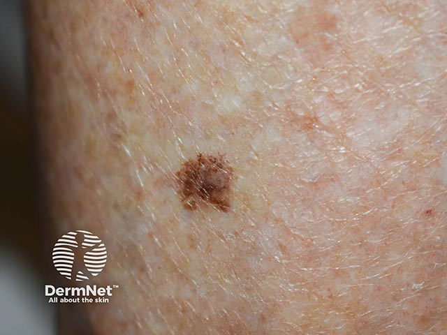
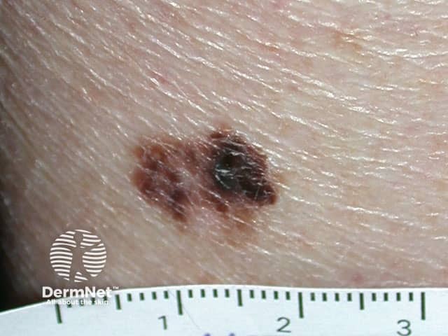
An irregularly marginated variably pigmented changing melanocytic lesion - it was a 0.5mm Breslow thickness melanoma
(SSM-patient10)
Dermoscopy of an irregularly pigmented melanocytic lesion - histology showed a thin invasive melanoma (SSM-patient10)
A changing irregularly pigmented lesion with a new nodule at the inferior pole - a malignant melanoma (SSM-patient11)
Go to the superficial spreading melanoma topic page
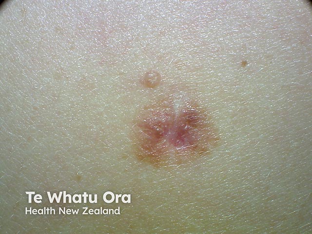
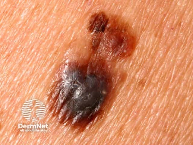
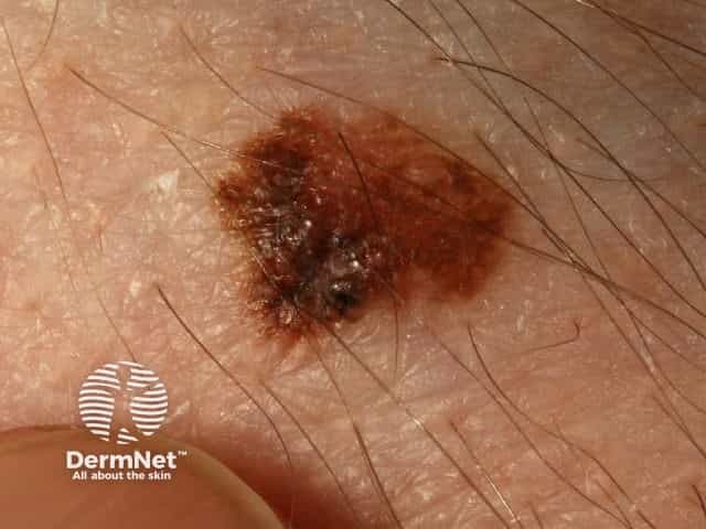
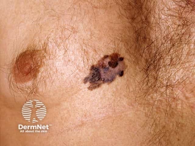
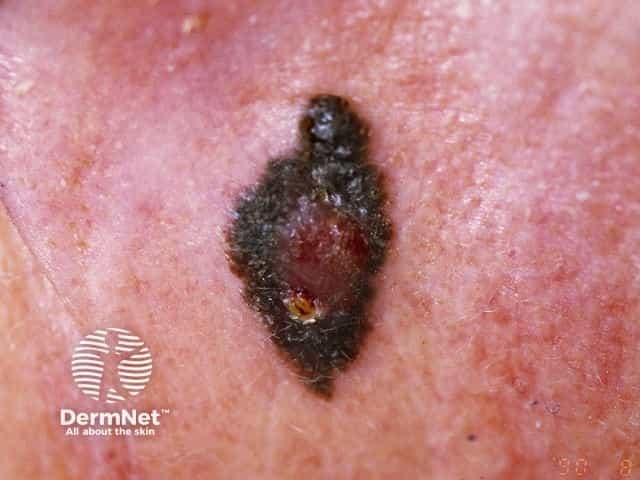
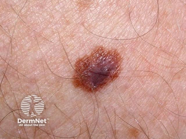
Superficial spreading melanoma macro images
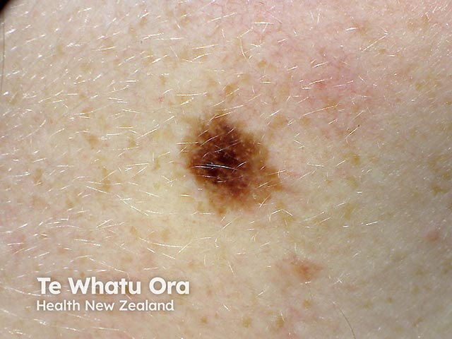
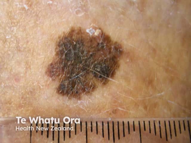
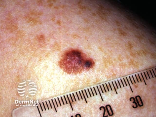
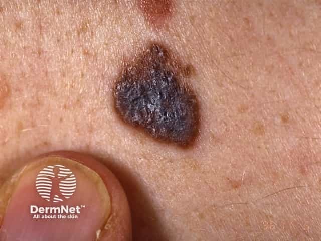
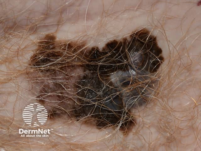
0.8 mm lentigo maligna melanoma
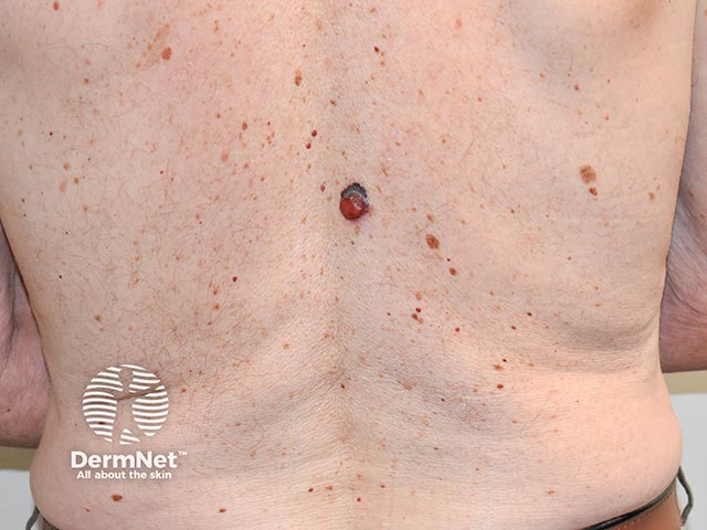
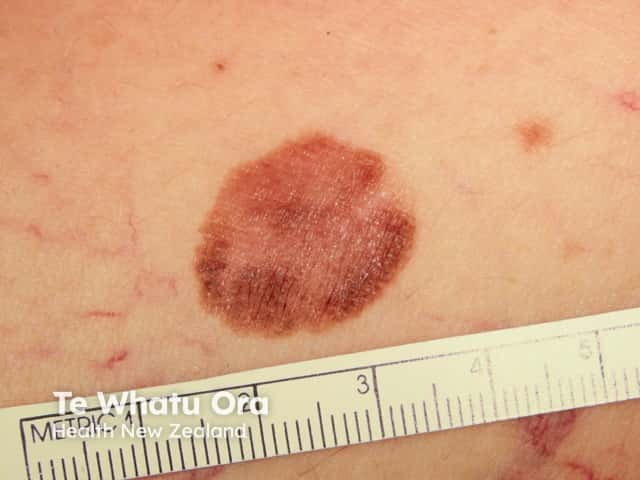
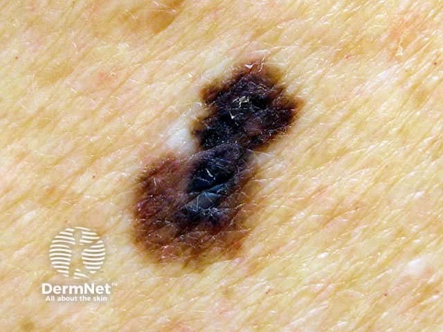
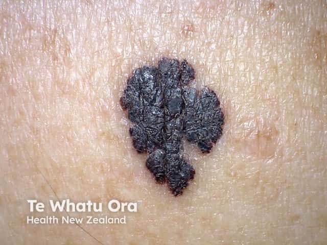
Superficial spreading melanoma
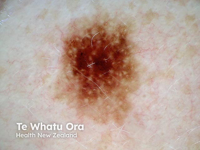
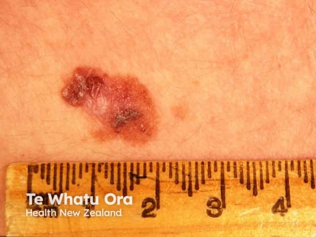
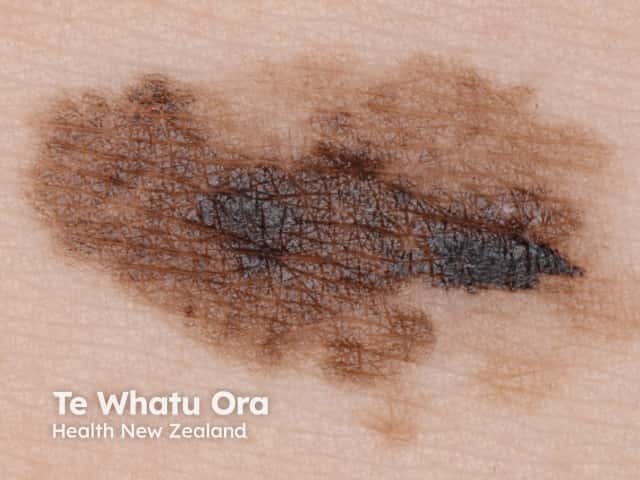
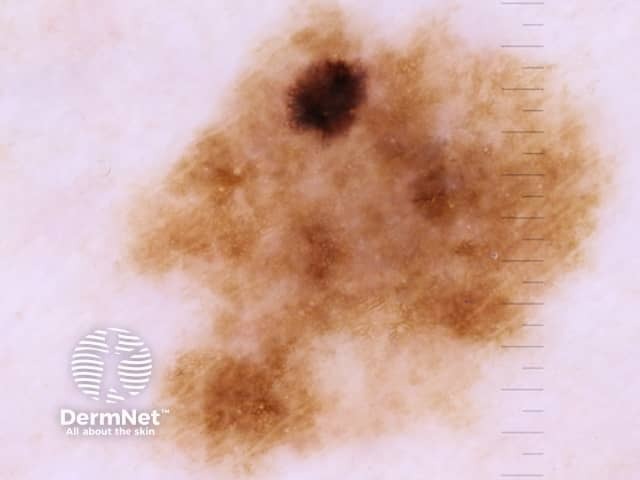
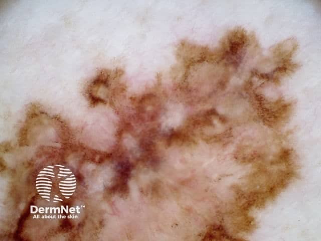
SSM with marked regression
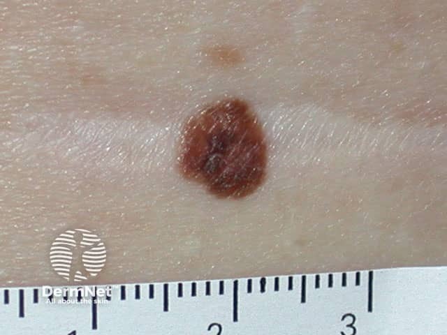
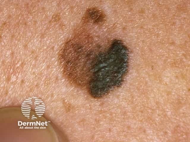
Superficial spreading melanoma
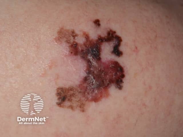
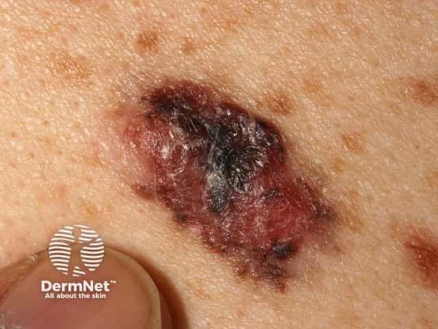
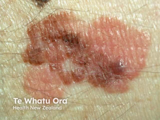
A superficial malignant melanoma - irregular and notched margin, variable and irregular pigmentation in an itchy and enlarging pigmented lesion
Go to the superficial spreading melanoma topic page
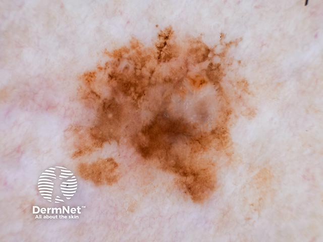
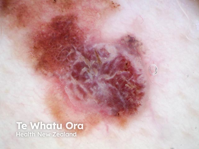
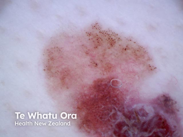
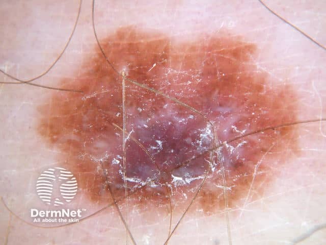
Dermoscopy images of superficial spreading melanoma
Superficial spreading melanoma dermoscopy
Superficial spreading melanoma dermoscopy
Superficial spreading melanoma dermoscopy
Superficial spreading melanoma dermoscopy
Amelanotic superficial spreading melanoma dermoscopy
Amelanotic superficial spreading melanoma dermoscopy
Superficial spreading melanoma dermoscopy in teenager
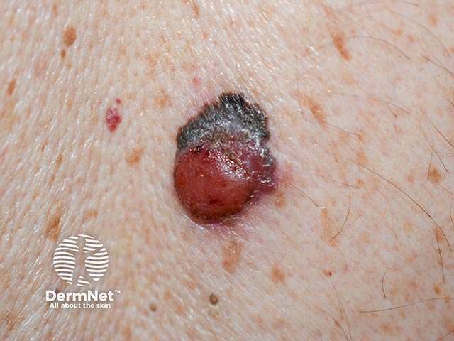
Dermoscopy of nodular melanoma arising within a superficial spreading melanoma
Superficial spreading melanoma, Breslow 0.3 mm
Go to the superficial spreading melanoma topic page
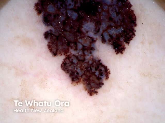
Dermoscopy images of superficial spreading melanoma
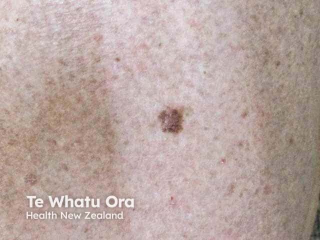
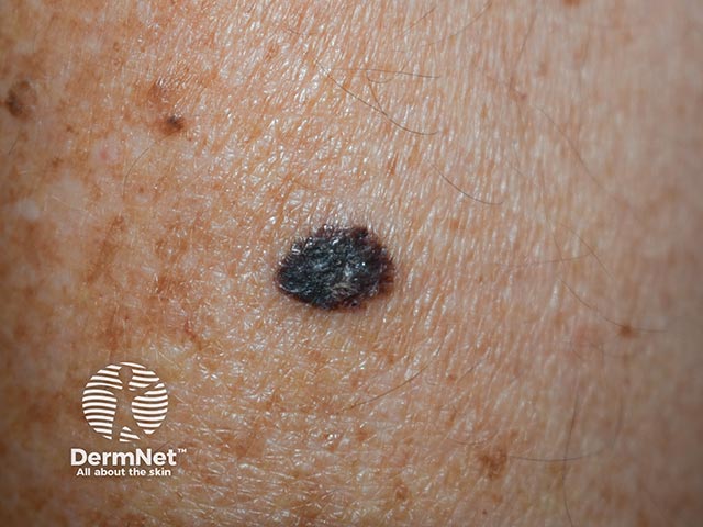
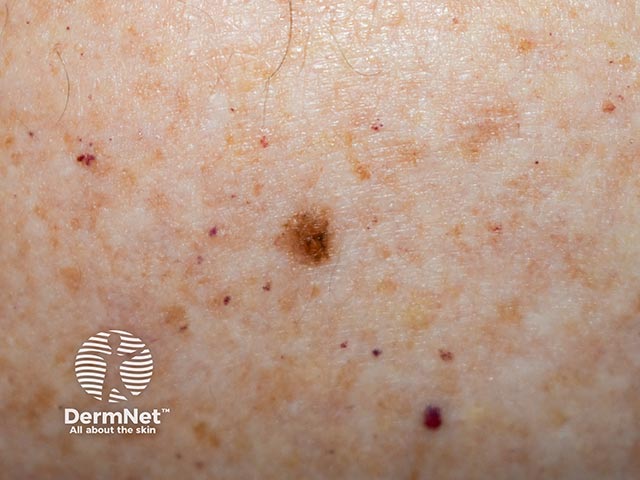
Macro image of superficial spreading melanoma 1
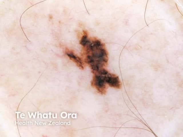
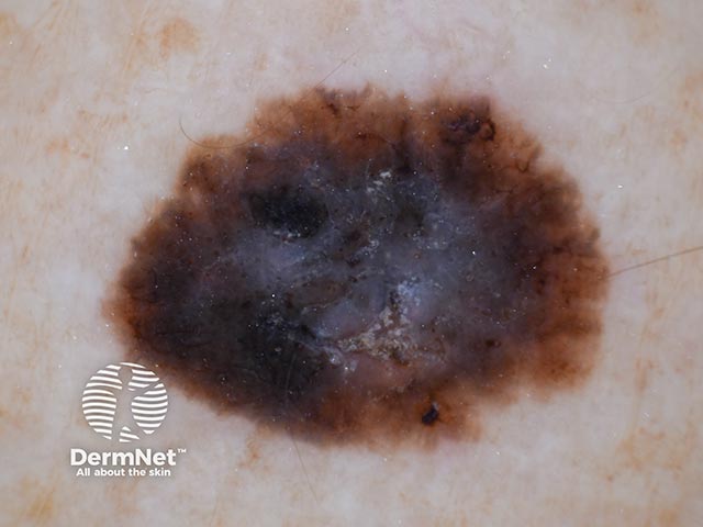
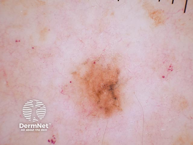
Dermoscopic image of superficial spreading melanoma 1
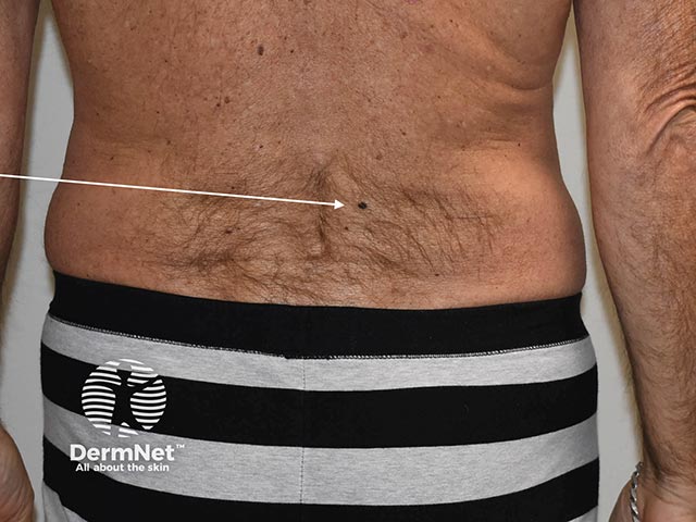
Macro image of superficial spreading melanoma 2
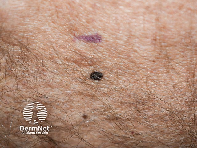
Dermoscopic image of superficial spreading melanoma 2
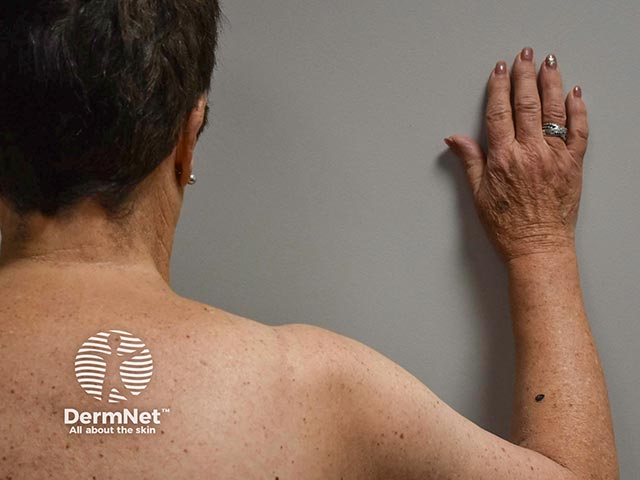
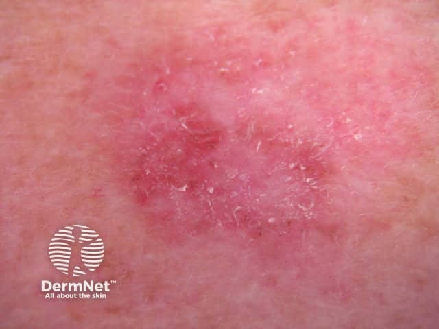
Macro image of superficial melanoma 3
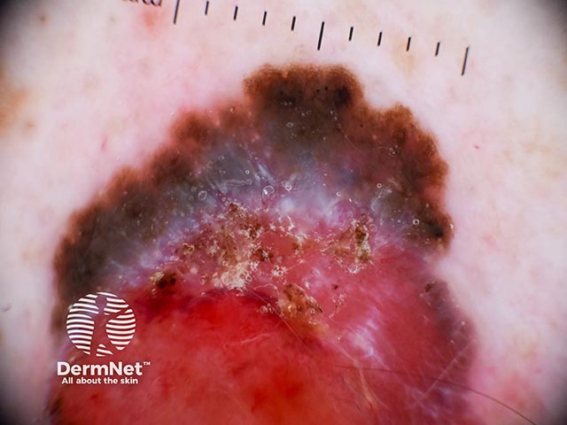
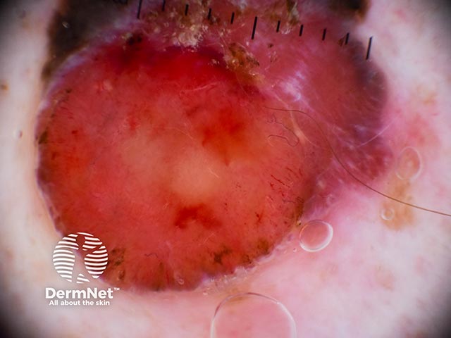
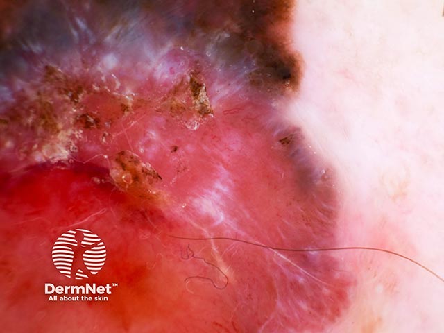
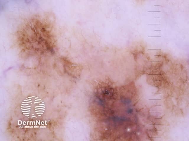
Dermoscopic image of superficial spreading melanoma 3 (top right half of lesion)
Dermoscopic image of superficial spreading melanoma 3 (bottom left half of lesion)
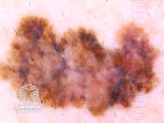
Macro image of superficial spreading melanoma 4
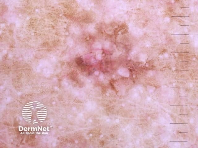
Dermoscopic image of superficial spreading melanoma 4 (top half of lesion)
Dermoscopic image of superficial spreading melanoma 4 (bottom half of lesion)
Macro image of superficial spreading melanoma 5
Dermoscopic image of superficial spreading melanoma 5
Macro image of superficial spreading melanoma 6
Dermoscopic image of superficial spreading melanoma 6
Macro image of superficial spreading melanoma 7
Dermoscopic image of superficial spreading melanoma 7
Macro image of superficial spreading melanoma 8
Dermoscopic image of superficial spreading melanoma 8
Macro image of superficial spreading melanoma 9
Dermoscopic image of superficial spreading melanoma 9
Go to the superficial spreading melanoma topic page