Main menu
Common skin conditions

NEWS
Join DermNet PRO
Read more
Quick links
Lesions (cancerous) Diagnosis and testing
Author: Anoma Ranaweera B.V. Sc; PhD (Clinical Biochemistry, University of Liverpool, UK); Chief Editor: Dr Amanda Oakley, Dermatologist, Hamilton, New Zealand, June 2014.
Introduction
Introduction - electrical impedance spectroscopy
How it works
Advantages
Clinical evidence and efficacy
Clinical use
Indications
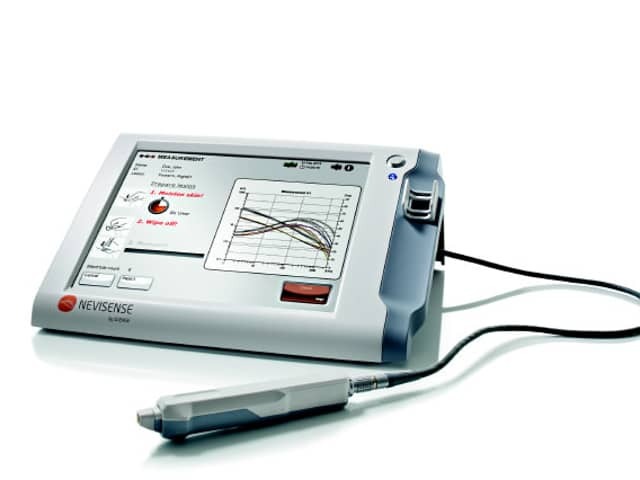
Electrical impedance spectroscopy (EIS) is used by a point-of-care portable device called Nevisense™ (Scibase AB, Stockholm, Sweden) to objectively analyse lesions with suspicion of melanoma.

Nevisense procedure

Nevisense device
*Images supplied by Scibase
Visual examination usually is sufficient when identifying most types of skin lesion. However, when it comes to atypical lesions, a clinical diagnosis based on only a visual examination may pose a challenge, leading to unnecessary excisions or even missed malignancies.
Dermatoscopy uses a hand-held skin surface microscope to examine skin lesions. In expert hands increases the chance of correct diagnosis, but requires training and considerable experience.
In the spectrometric analysis of skin lesions, a computer software program is used that calculates and extracts information about the cells and structures of the skin. The method uses a light beam that penetrates beneath the skin surface. Light images taken with a digital camera or hand-held scanner are fed into a computer for detailed analysis.
In electrical impedance spectroscopy, the varying electrical properties of human tissue are used to categorise cellular structures and thereby detect malignancies.
SciBase Electronic Impedance Spectroscopy is a patented technology developed at the Karolinska Institute in Stockholm, Sweden.
Electrical impedance spectroscopy analysis complements the physicians' visual examinations, particularly in cases of cutaneous lesions with unclear clinical signs of melanoma. Benefits include: