Introduction
Dermatoscopic features of pigmented lesions
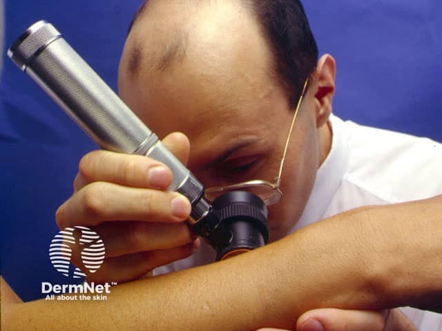
Dermatoscopes
Dermoscopy or dermatoscopy refers to the examination of the skin using skin surface microscopy, and is also called ‘epiluminoscopy’ and ‘epiluminescent microscopy’. Derm(at)oscopy is mainly used to evaluate pigmented skin lesions. In experienced hands it can make it easier to diagnose melanoma.
Dermoscopy requires a high quality magnifying lens and a powerful lighting system (a dermatoscope). See below for more information on dermatoscopes.
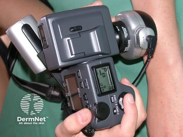
Computer software can be used to archive dermoscopy images and allow expert diagnosis and reporting (mole mapping). Smart programs may aid in diagnosis by comparing the new image with stored cases with typical features of benign and malignant pigmented skin lesions.
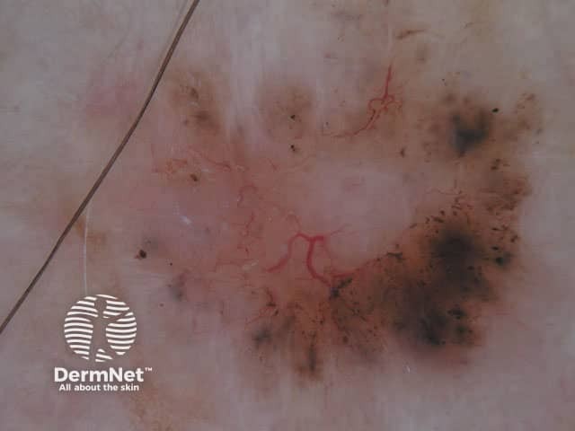
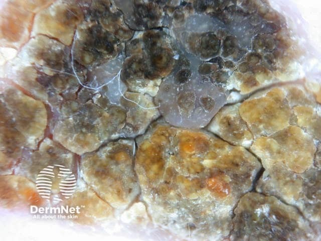
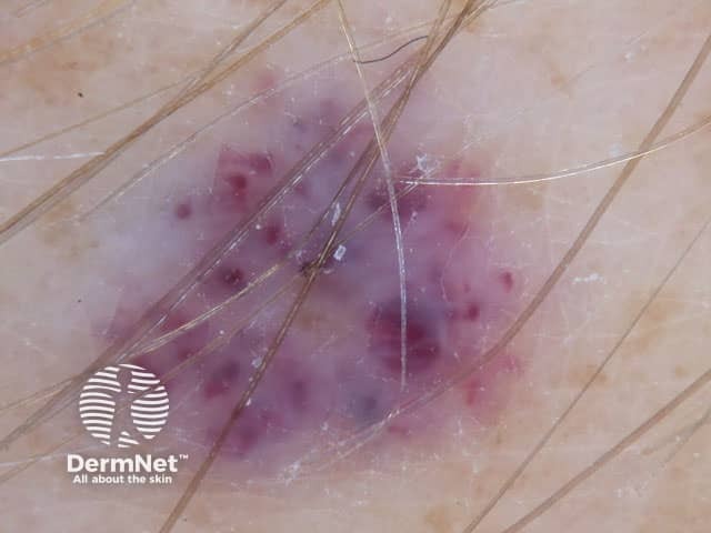
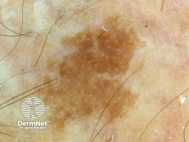
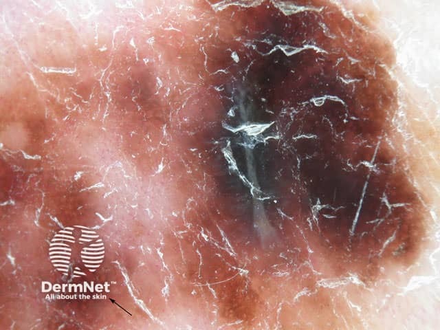
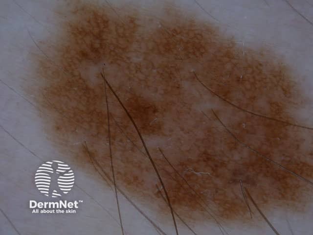
Using dermoscopy, the pigmentation of the lesion is evaluated in terms of colour(s) and structure.
Colours found in pigmented skin lesions include black, brown, red, blue, grey, yellow and white.
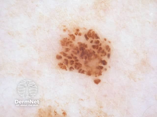
Characteristics of the dermatoscopic structure of the skin lesions include:
There are specific dermoscopic patterns that aid in the diagnosis of the following pigmented skin lesions:
Dermoscopy may also be used for detailed skin surface examination in some other circumstances, for example:
Used widely by dermatologists, plastic surgeons, and general practitioners, dermatoscopes allow the user to perform skin surface microscopy. This enables the practitioner to examine skin structures and patterns.
There are several different dermatoscopes and these are either conventional standalone devices, or smartphone attachable. When choosing a dermatoscope, there are some important things to consider:
See also our Dermatoscope comparative image gallery.

