DermNet provides Google Translate, a free machine translation service. Note that this may not provide an exact translation in all languages

Translate

Infantile haemangioma: Definition and pathogenesis

Authors: Dr Amanda Oakley, Dermatologist, Hamilton, New Zealand, 1997. Updated 2012. Revised and updated Dr Arun Gangakhedkar, Paediatrician, Waitemata DHB, Auckland, New Zealand. Copy edited by Gus Mitchell. January 2021.


toc-icon

What is infantile haemangioma?

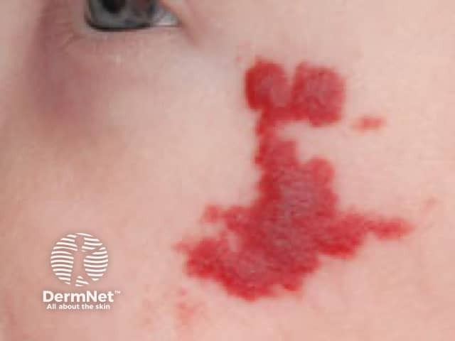
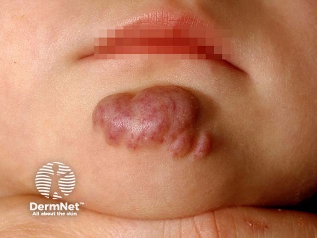
Infantile haemangioma, also known as a strawberry naevus, is the most common benign vascular skin tumour in children. It is noticed in the first few weeks of life.

Infantile haemangiomas on the face

Who gets infantile haemangioma?

Infantile haemangioma is found in 1–2% of newborns, and up to 10–12% at 1 year of age in Caucasian infants. It is less frequent in non-Caucasian populations. Girls are more commonly affected than boys (3:1).

Risk factors for developing infantile haemangioma include:

  • Low birth weight increases the risk by 40% for every 500g decrease in birth weight. Infantile haemangioma affects 1 in 4 preterm infants weighing under 1000g.
  • Maternal factors carrying increased risk include: advanced maternal age, subfertility treatment, multiple pregnancy (eg, twins, triplets), pre-eclampsia, and placenta praevia.

What causes infantile haemangioma?

A placental origin for infantile haemangioma is suggested due to GLUT1 protein expression. Fetal vascular origins are proposed due to the presence of primitive marker CD133 from the fetal cardinal vein.

Defective vascular stem cell regulation involving endothelial progenitor cells (EPC), or extrinsic factors such as hypoxia and developmental vascular field disturbances can influence the development of vascular anomalies.

What are the clinical features of infantile haemangioma?

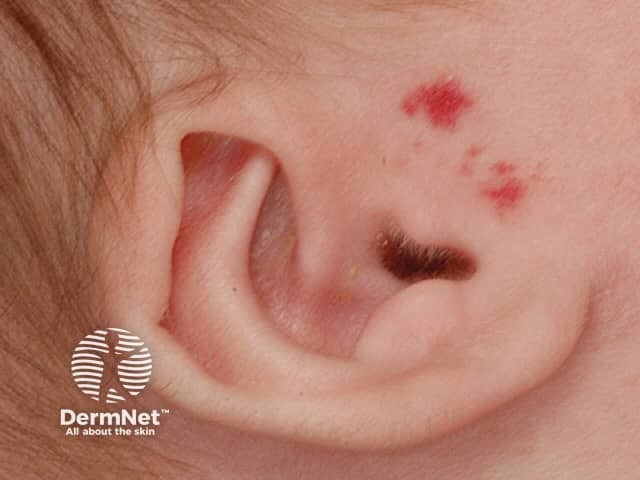
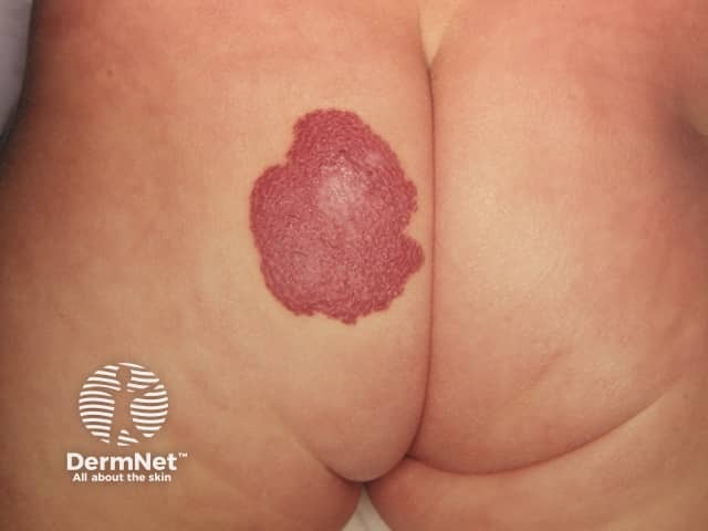
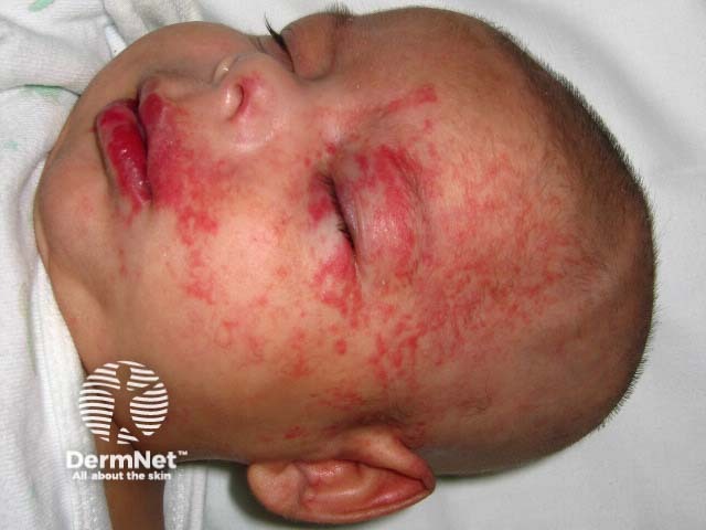
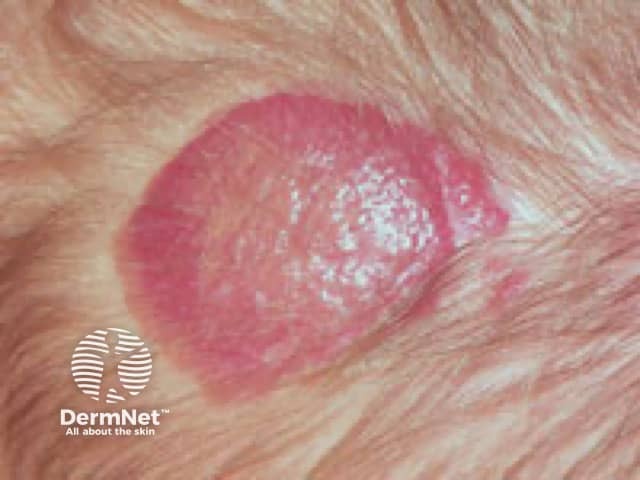
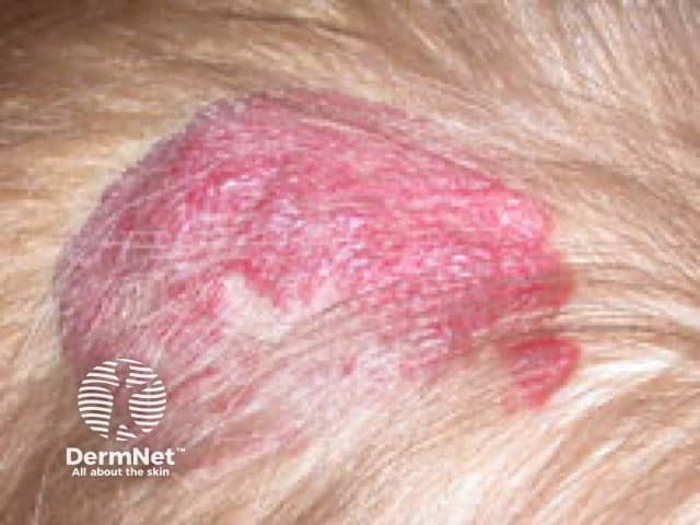
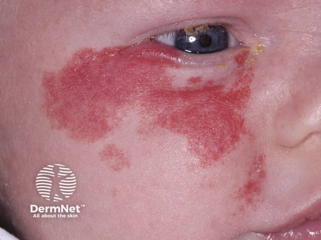
Infantile haemangiomas are typically solitary and most are located on the head and neck region (60%). Involvement of internal organs is usually indicated by multiple cutaneous lesions. The clinical features are determined by the depth of the lesion, distribution pattern, and phase of growth.

Depth

  • Superficial lesions in the upper dermis are bright red, non-pulsatile, non-tender papules and plaques, and are warm to touch.
  • Deep lesions in the deeper dermis and subcutaneous tissue were previously called cavernous haemangioma. They are flesh-coloured or blue, non-pulsatile, non-tender masses, and are warm to touch. Compared to the superficial lesions, these tend to appear later and have a late growth phase.
  • Mixed: features of both superficial and deep forms.

Infantile haemangiomas classified by depth

Distribution pattern

  • Focal (77%) — solitary, discrete, circular, or plaque-like lesion.
  • Segmental (18%) — plaques in an anatomic or developmental distribution commonly associated with local complications or structural anomalies and continued growth beyond 6 and occasionally 9 months of age (3%).
  • Multifocal (3%) — several discrete localised lesions distributed over more than one anatomic site.
  • Indeterminate — the lesions are not definitively focal or segmental.

Distribution patterns of infantile haemangioma

Growth phases

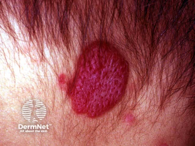
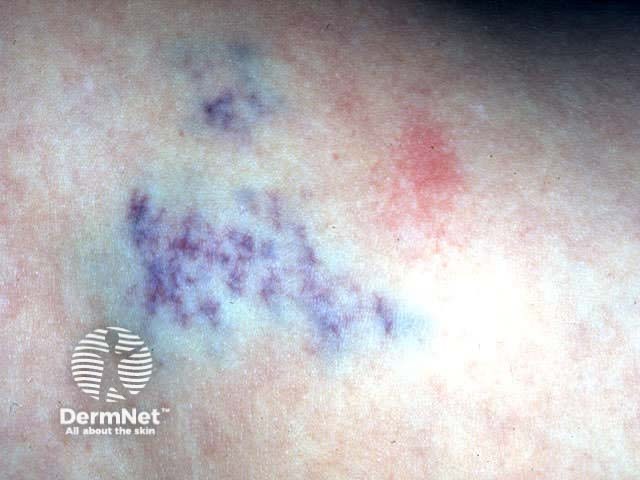
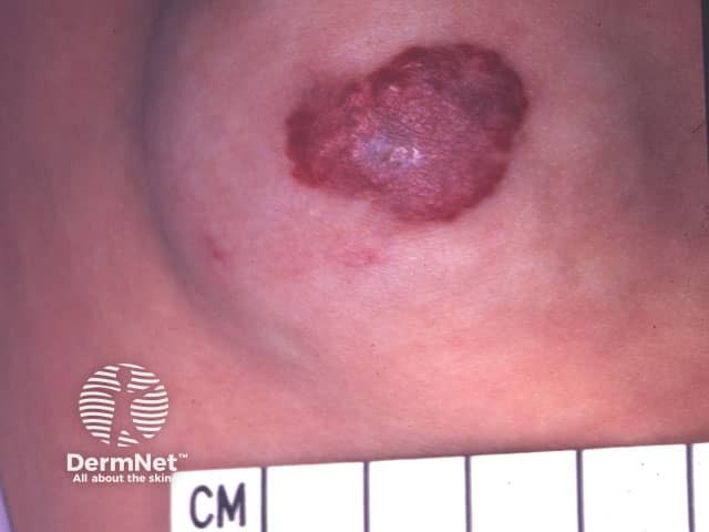
Most infantile haemangiomas show rapid growth in the first 3 months with a growth arrest by about 5 months of age, receding subsequently over several years. The progression may be described in different morphological phases.

  • Prodromal, nascent, or premonitory phase: A precursor lesion can be a circumscribed erythematous macule or paradoxical ischaemic patch (herald patch); have telangiectasia with an ischaemic halo, anaemic or blue-tinged bruise-like discolouration; or can be a blurred swelling.
  • Initial phase (1–3 weeks age): Loss of typical skin texture with increasing thickness and an indurated appearance by 2–4 weeks of age.
  • Proliferation phase (1–3 months age): Firm, non-compressible, bright red with infiltration and a mostly exophytic surface growth.
  • Maturation phase (plateau phase): Raised, bumpy, crimson red lesions reach maximum size by the first birthday. Regression and proliferation may be seen in same lesion.
  • Regression or involution phase (several years): The bright red colour changes to a dull grey surface associated with flattening, softening, and shrinking from the centre towards the periphery. While a grey stage is suggestive of involutional change, a white infantile haemangioma may be a sign of impending ulceration. The most rapid phase of involution is seen between 1–4 years and approximately 50% of lesions involute by 5 years, 70% by 7 years, and 90% by 9 years of age.

Scalp infantile haemangioma, various growth phases

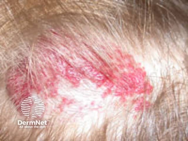
Infantile haemangioma with minimal or arrested growth (IH-MAG) has an abortive or minimal growth in 25% of the lesion’s surface area, appearing as telangiectatic patches with or without papules, lacking a significant proliferative phase. This may be mistaken for a port-wine stain [see Capillary vascular malformation]. Occasionally these can be segmental with syndromic associations. Two-thirds of these lesions are seen over lower limbs.

How is infantile haemangioma diagnosed?

Infantile haemangioma is usually a clinical diagnosis and investigations are not routinely indicated. Investigations may be considered if the diagnosis is uncertain, to define extent and associations, or monitor response to therapy.

Tests may include:

  • Doppler ultrasonography
  • MRI for suspected anatomical associations such as PHACE, SACRAL, LUMBAR and PELVIS syndromes
  • Laryngoscopy to investigate stridor with or without beard lesions
  • For visceral involvement — thyroid and liver function tests, ultrasound of abdomen, MRI or ultrasound of the head, or echocardiogram
  • Skin biopsy shows uniform vessel morphology with an outer layer of pericytes and inner lining of endothelial cells containing typical microtubular Weibel-Palade bodies
  • Immunohistochemical staining for GLUT1 confirms a diagnosis of infantile haemangioma, although GLUT1 is also positive in angiosarcoma, epithelioid endothelioma, angiokeratoma, and verrucous haemangioma. Staining is negative in congenital haemangioma and other vascular malformations.

What is the differential diagnosis for infantile haemangioma?

  • Vascular malformations — are present at birth and persist with no proliferative or involutional phase.
  • Vascular tumours — include congenital haemangioma, pyogenic granuloma, tufted angioma.
  • Locally aggressive tumours — Kaposiform haemangioendothelioma, Kaposi sarcoma.
  • Malignant tumour — angiosarcoma.

Differential diagnosis of infantile haemangioma

[see also Infantile haemangioma: Complications and treatment]


Bibliography

  • Darrow DH, Greene AK, Mancini AJ, Nopper AJ. Diagnosis and management of infantile hemangioma. Pediatrics. 2015;136(4):e1060-104. doi:10.1542/peds.2015-2485. PubMed
  • Gelmetti C. Vascular birthmarks: A hidden world behind a word. Indian J Paediatr Dermatol. 2018;19:1–8. doi:10.4103/ijpd.IJPD_124_17. Journal
  • Greenberger S, Bischoff J. Pathogenesis of infantile haemangioma. Br J Dermatol. 2013;169(1):12–9. doi:10.1111/bjd.12435. PubMed
  • Harter N, Mancini AJ. Diagnosis and management of infantile hemangiomas in the neonate. Pediatr Clin North Am. 2019;66(2):437–59. doi:10.1016/j.pcl.2018.12.011. PubMed
  • Higgins EM, Glover MT. Dermatoses and haemangiomas of infancy. In: Griffiths C, Barker J, Bleiker T, Chalmers R, Creamer D (eds). Rook’s Textbook of Dermatology, 9th edn. Wiley Blackwell, 2016: 117.16-23.
  • Soliman YS, Khachemoune A. Infantile hemangiomas: our current understanding and treatment options. Dermatol Online J. 2018;24(9):13030/qt5jt8q9km. PubMed

On DermNet

Other websites

Books about skin diseases


Related information

Sign up to the newsletter