Main menu
Common skin conditions

NEWS
Join DermNet PRO
Read more
Quick links
Dermoscopy is very useful for the diagnosis of pigmented lesions.
This quiz requires knowledge of conventional dermoscopic pattern analysis. It was originally in the form of a column written by Dr Amanda Oakley and published by New Zealand Doctor in February 2010. It was revised and prepared for DermNet by Niket Shah, final year medical student, University of Otago, in May 2020.
For each of the twelve cases, study the image(s) and then answer the questions. You can click on the image to view a larger version if required. Each case should take approximately 2 minutes to complete.

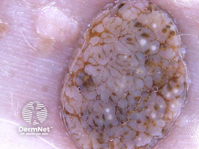
Name the dermoscopic pattern.
This lesion has a nonspecific pattern.
There are milia-like cysts (white clods), irregular crypts (orange and brown clods) and cerebriform structures i.e., ridges and furrows (thick curved and parallel lines). There are no signs of a melanocytic lesion as there is no pigment network or aggregated globules.
What is the diagnosis?
Seborrhoeic keratosis