This quiz tests your ability to recognise different causes of purpura.
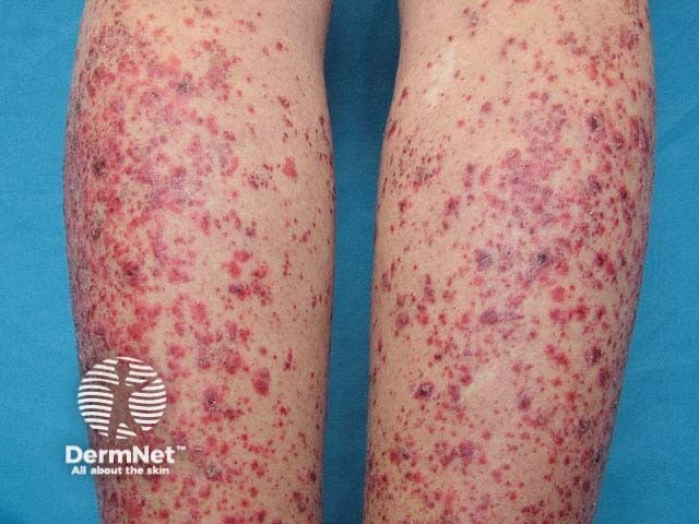
Purpura is due to haemorrhage from small blood vessels, and there is as wide differential diagnosis. Petechiae are small, purpuric lesions up to 2mm across, and ecchymoses or bruises are larger. Extravasated blood usually breaks down and changes colour over a few weeks from purple, orange, brown and even blue and green.
Purpura may arise because of platelet destruction or malfunction, as in primary or secondary thrombocytopaenic purpura. This may result in petechiae, ecchymoses and external bleeding.
Severe coagulation disorder associated with disseminated intravascular coagulation may result in purpura fulminans with generalised, large spontaneous ecchymoses and external bleeding. Petechiae do not feature. The purpura results from thrombotic vaso-occlusion associated with deficiency of protein C, protein S and antithrombin III.
Vascular damage may result in persistent and localised purpura, often with an erythematous and palpable inflammatory component. Ecchymoses and external bleeding are uncommon. Purpura may be due to vessel wall deposition of metabolic substances, such as amyloid in systemic amyloidosis, or calcium in calciphylaxis. Deficiency of ascorbic acid results in scurvy, in which petechiae are especially seen surrounding twisted hairs but may result in large areas of purpura and ecchymoses. Purpura may be caused by vaso-occlusion by emboli, thrombi, or fibrointimal hyperplasia (endarteritis obliterans). Clinical presentations include coumadin necrosis, antiphospholipid antibody syndrome, cardiac myxoma and radiation arteritis.
Inflammatory reactions may be due to large vessel disease (polyarteritis nodosa), small vessel vasculitis, capillaritis, or localised insect bite reactions.
For each of the ten cases, study the image(s) and then answer the questions. You can click on the image to view a larger version if required.
Each case should take approximately 2 minutes to complete. There is a list of suggested further reading material at the end of the quiz.
When you finish the quiz, you can download a certificate.
Make a diagnosis.
What features distinguish this condition from the others?
Finished!
If you would like to receive a certificate of participation please enter your name below and click Download.

