White skin lesions may be quite unsightly, particularly in dark skin. Some are symptomatic, and others may be seen in association with serious skin diseases. At times the different causes can be hard to distinguish, even by experts.
It is important to distinguish pallor or hypopigmentation (decreased skin colour) from leukoderma or achromia (no pigment at all). Take a careful history: When and where was the first patch? Was the condition present at birth? Family history? Did it follow an inflammatory skin disorder or injury? Is it continuing to spread? Are there symptoms, including decreased sensation? What was the effect of treatment?
When examining the skin, note the site(s), size, morphology and distribution. Is/are the lesion/s patches (thin), plaques (thick) or indurated (firm); smooth, dry or scaly? What does neighbouring skin look like?
For each of the twelve cases, study the image(s) and then answer the questions. You can click on the image to view a larger version if required.
Each case should take approximately 2 minutes to complete. There is a list of suggested further reading material at the end of the quiz.
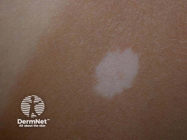
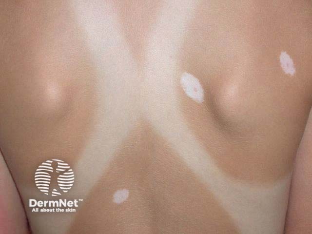
Name the skin condition causing the white mark/s.
What is the cause?

