Introduction Clinical features Dermoscopic features BRAAFF algorithm Differential diagnoses Histology
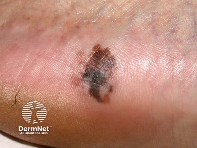
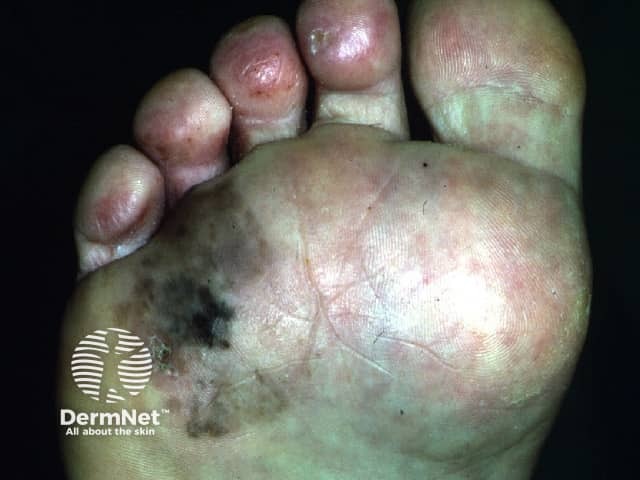
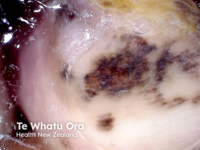
Acral lentiginous melanoma is a type of melanoma arising on the palms of the hands, soles of the feet, or the underside of fingers or toes.
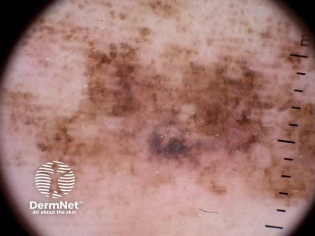
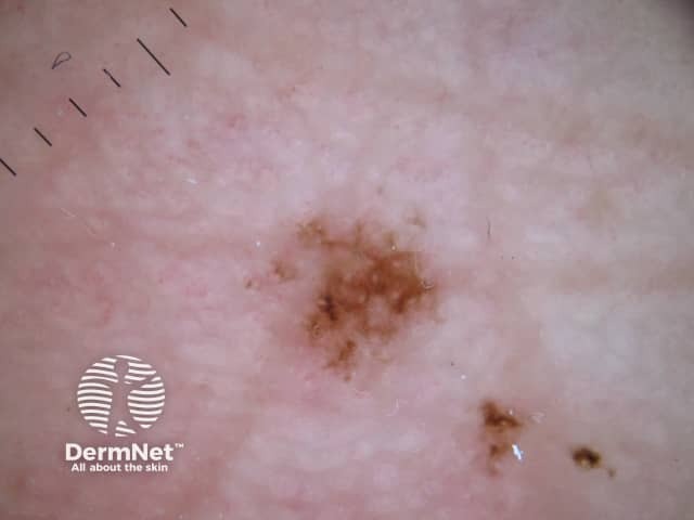
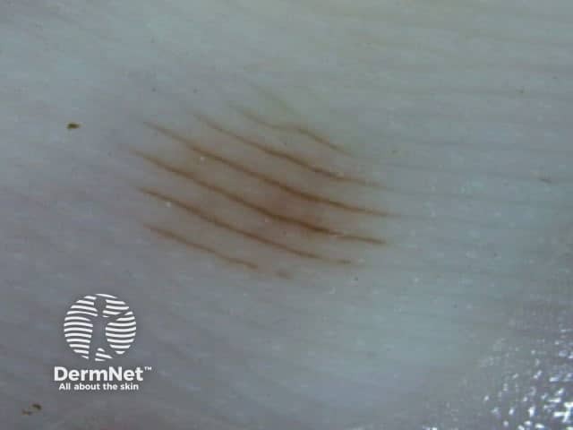
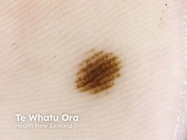
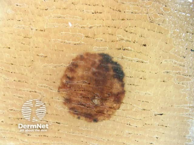
Acral lentiginous melanoma starts as a slowly enlarging flat patch of discoloured skin.
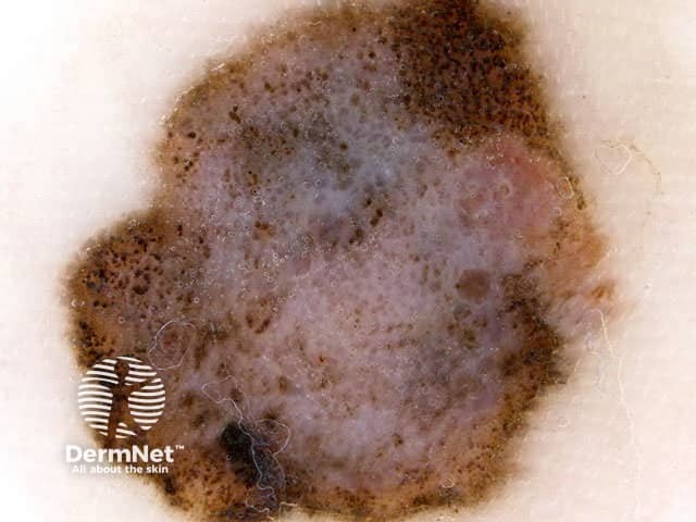
The characteristics of acral lentiginous melanoma include:
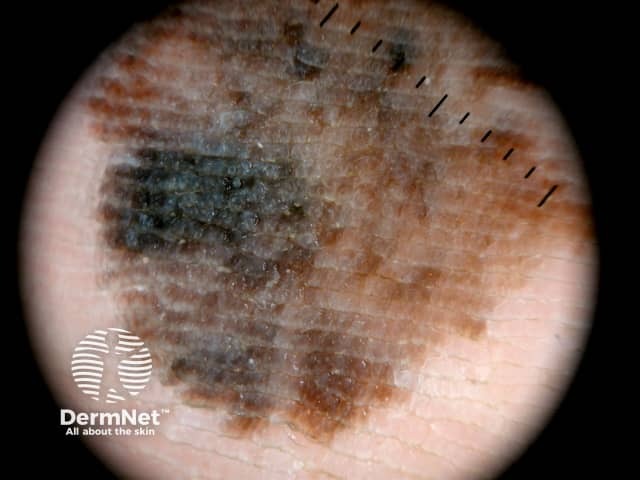
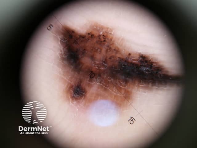
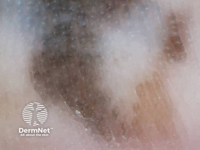
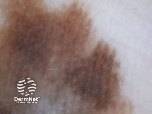
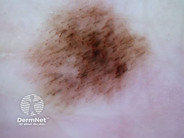
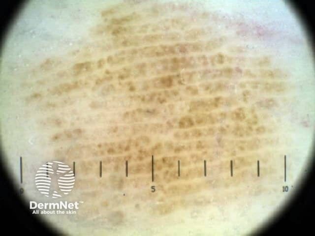
The most frequently observed dermoscopic features of acral lentiginous melanoma are:

In this algorithm, one point is required to make the diagnosis of melanoma [1].
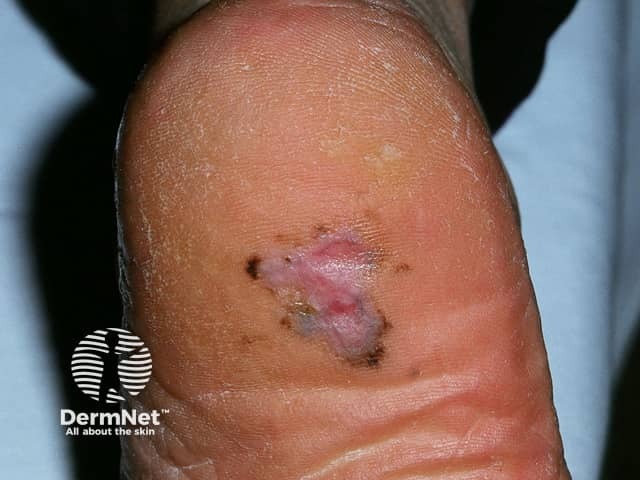
It can be clinically and dermoscopically challenging to make the correct diagnosis in pigmented acral lesions. The following are differential diagnoses of acral lentiginous melanoma.
These dermoscopic patterns are termed the parallel ridge pattern and the parallel furrow pattern, respectively. The sensitivity and specificity of the parallel ridge pattern in diagnosing early acral melanoma is 86% and 99%, respectively. However, dermoscopic features in acral naevus (especially congenital acral naevus) sometimes mimic the parallel ridge pattern [2].
The presence of a red-black homogeneous pigmentation, often combined with satellite globules is most likely indicative of haemorrhage. A positive scratch test (where removing a sample of the surface of the skin surface reveals blood) may be considered as an additional diagnostic clue to differentiate from melanoma [3].
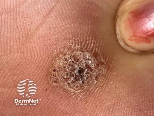
Irregular pigmentation at the edge of a warty nodule could be indicative of melanoma [4].
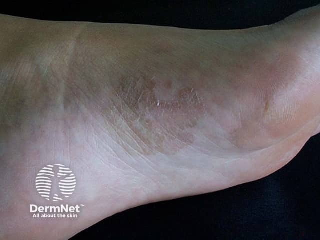
Tinea nigra, a superficial fungal infection caused by Phaeoannellomyces werneckii, presents as a hyperpigmented, nonscaling macule of variable size and shape. Typically lacking induration, erythema, or pruritus, these 'ink spot' lesions may resemble junctional naevus or melanoma [5].
The pathological diagnosis of melanoma can be very difficult. Histological features of acral lentiginous melanoma include an asymmetrical proliferation of melanocytes at the dermo-epidermal junction.
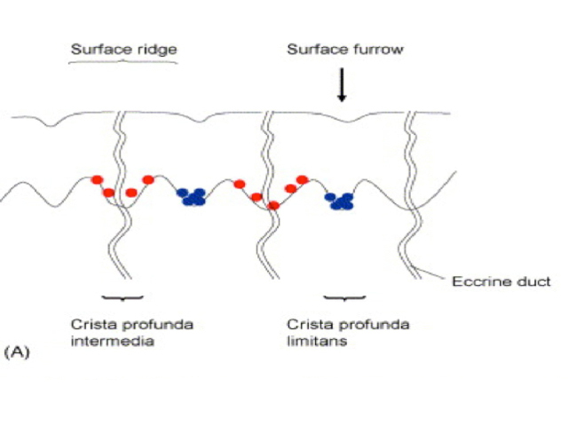
Preferential proliferation patterns of early melanoma cells and naevus cells in acral volar skin. Early melanoma cells (red dots in the diagram below) are mainly found as individual cells in the crista profunda intermedia under the surface ridge. In contrast, nests of acral naevus cells (blue dots in the diagram) are seen in the crista profunda limitans under the surface furrow [6].