Introduction
Histology
Special studies
Differential diagnoses
Angiokeratoma presents as a blood-filled papule which may bleed following trauma. There are a range of clinical presentations ranging from isolated lesions of little consequence to widespread lesions associated with Fabry disease.
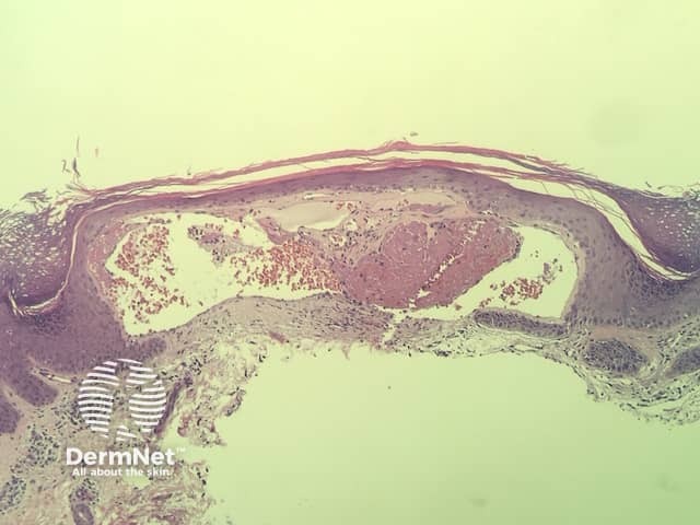
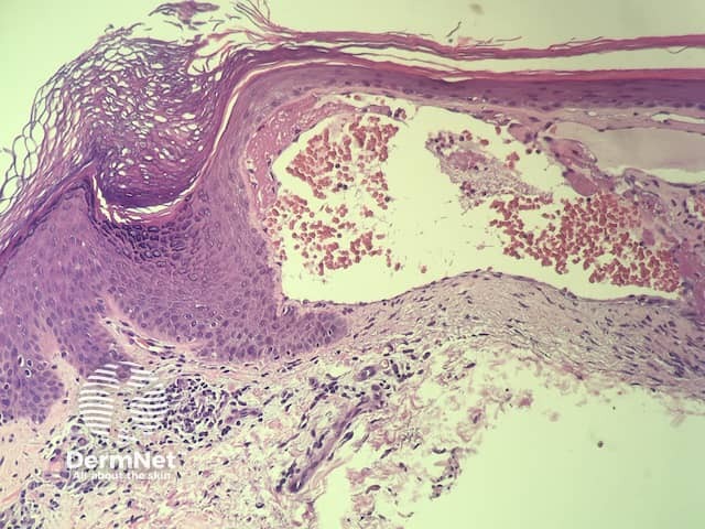
In angiokeratoma, the histopathology shows a vascular lesion in the superficial dermis which extends into the epidermis. There is epidermal hyperplasia and papillomatosis. There may be intravascular thrombosis (figures 1,2).
None are generally needed.
Other diagnoses to be considered include:

