Introduction Histology Special studies Differential diagnoses
Angiomyofibroblastoma is a benign soft tissue tumour of the external genitalia.
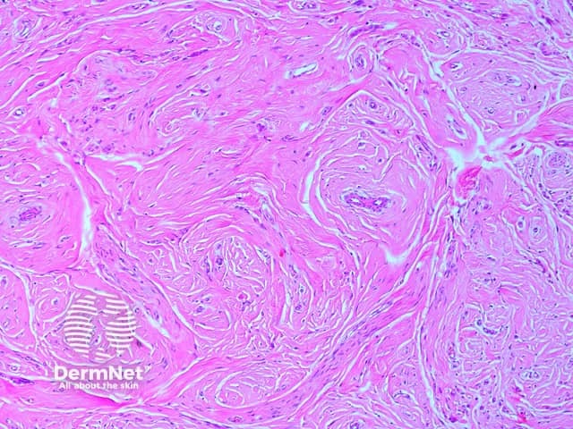
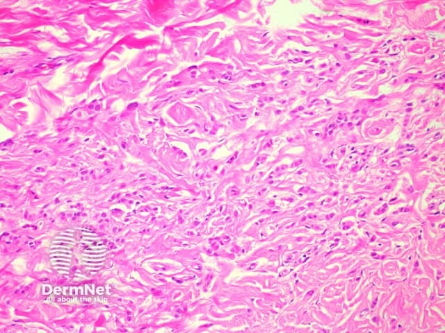
Microscopic examination of angiomyofibroblastoma demonstrates a well-circumscribed tumour without a capsule and with variable cellularity. Hypocellular areas are collagenous (figure 1) or myxoid with some small-medium sized vessels. Hypercellular areas, located predominately around the vessels, contain characteristic plump round/oval shaped cells with relatively uniform nuclei without remarkable atypia (figure 2). Occasional multinucleated and binucleated cells may be seen.
The angiomyofibroblastoma tumour cells are positive with immunohistochemistry for actin. Oestrogen and progesterone markers may also be positive.
Aggressive angiomyxoma – These are infiltrative tumours and usually much larger (more than 5 cm). Tumours have been described with mixed features of angiomyofibroblastoma and aggressive angiomyxoma, which raises the possibility that these tumours are related.

