

Annular lichenoid dermatitis of youth — extra information
Introduction Demographics Causes Clinical features Diagnosis Differential diagnoses Treatment Outcome
What is annular lichenoid dermatitis of youth?
Annular lichenoid dermatitis of youth is a rare skin condition that was first described in 2003 [1]. It is characterised by sharply demarcated annular erythematous macules and papules with central hypopigmentation [2].
Who gets annular lichenoid dermatitis of youth?
Annular lichenoid dermatitis of youth was first described in young healthy individuals from the Mediterranean [1]. The mean age of patients affected by this skin condition is 10 years [2]. Cases in adults and older people have been reported from central Europe, the USA, and Japan [3,4].
What causes annular lichenoid dermatitis of youth?
An exact cause for annular lichenoid dermatitis of youth is yet to be discovered. It is believed to share pathogenic factors with other lichenoid dermatoses, like lichen planus, graft versus host disease, and lichenoid drug eruption, which involve a T-cell-mediated response and subsequent apoptosis of basal keratinocytes [2]. The reasons why the rete ridges are targeted in annular lichenoid dermatitis are currently unknown.
What are the clinical features of annular lichenoid dermatitis of youth?
Annular lichenoid dermatitis typically presents as asymptomatic rounded, oval, or annular red-brown macules or patches. These start as erythematous macules with a raised border and develop central hypopigmentation as they gradually enlarge (Figure 1) [2]. The macules or patches are not itchy.
The lesions are found on the patient's trunk, groin, and less frequently, in the axillae [2]. The individual is otherwise well, with no systemic symptoms being seen.
Figure 1. Annular lichenoid dermatitis of youth
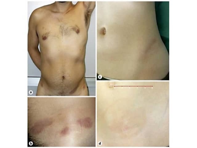
Credit: Prof Paolo Gisondi
How is annular lichenoid dermatitis of youth diagnosed?
Annular lichenoid dermatitis of youth is diagnosed on histopathology, with the presence of specific hallmark features. These features include:
- An interface dermatitis only affecting the tips of the rete ridges (Figure 2) [2]
- A loss of keratinocytes, resulting in blunting of the rete ridges, giving a squared-off appearance (Figure 2) [2].
Figure 2. Histopathological features of annular lichenoid dermatitis of youth
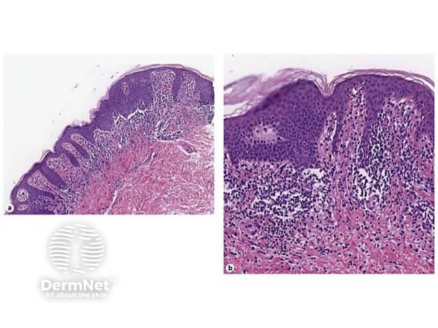
Credit: Prof Paolo Gisondi
What is the differential diagnosis for annular lichenoid dermatitis of youth?
Annular lichenoid dermatitis of youth may be clinically confused with morphoea, mycosis fungoides, and annular erythema.
- Morphoea is differentiated by its histopathological findings of an atrophic epidermis, thickened hyaline collagen, or loss of sweat glands and hair follicles.
- Hypopigmented mycosis fungoides is differentiated histopathologically by the infiltration of malignant T cells into the epidermis and by extensive spongiosis.
- Annular erythema typically self-resolves and is often scaly. The histopathological findings show an inflammatory cell infiltrate surrounding superficial vessels, known as the 'coat-sleeve anomaly'.
What is the treatment for annular lichenoid dermatitis of youth?
Transient remissions can be achieved in annular lichenoid dermatitis of youth with treatment with topical steroids, systemic steroids, topical tacrolimus, or narrowband ultraviolet B phototherapy [2].
The lesions can reappear once treatment has been ceased [2]. Spontaneous recovery without treatment can also occur, but this is uncommon.
What is the outcome for annular lichenoid dermatitis of youth?
Annular lichenoid dermatitis of youth slowly progresses, with no serious complications being reported to date. The duration of annular lichenoid dermatitis of youth varies. Out of 21 cases described from 2005 to 2013, most lesions were successfully treated with topical corticosteroids but these recurred once therapy was discontinued [2]. There have been a few cases that demonstrated complete remission after treatment cessation, and one case that showed spontaneous regression without any treatment [2].
References
- Annessi G, Paradisi M, Angelo C, Perez M, Puddu P, Girolomoni G. Annular lichenoid dermatitis of youth. J Am Acad Dermatol 2003; 49: 1029–36. DOI: 10.1016/S0190-9622(03)02147-9. PubMed
- Mercurio D, Gisondi P, Colato C, Schena D, Girolomoni G. Annular lichenoid dermatitis of youth: reports of six new cases with review of the literature. Dermatology 2015; 231: 195–200. DOI: 10.1159/000381705. Journal
- Huh A, Kanitakis J. Annular Lichenoid dermatosis of youth: report of the first Japanese cases and published work review. J Dermatol 2010; 37: 531–3. DOI: 10.1111/j.1346-8138.2009.00740.x. Journal
- Leger MC, Gonzalez ME, Meehan S, Schaffer JV. Annular lichenoid dermatitis of youth in an American boy. J Am Acad Dermatol 2013; 68: e155–6. DOI: 10.1016/j.jaad.2012.01.030. PubMed