Introduction Histology Special studies Differential diagnosis
Apocrine hidrocystomas are regarded by some authorities to represent cystic retention of apocrine glands. Others believe they are adenomatous lesions.
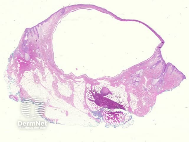
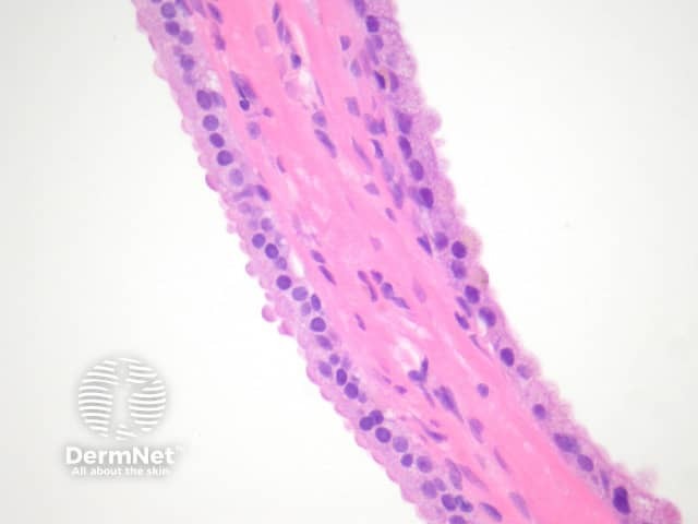
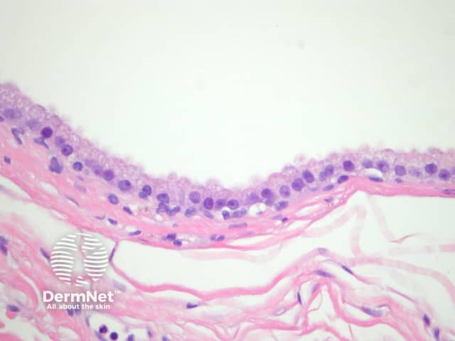
A cystic space occupies the dermis which may be unilocular (figure 1) or multilocular. The cystic space is lined by a bilaminar epithelium which has on its inner portion cells which are columnar, eosinophilic and show prominent luminal blebbing (‘apocrine snouts’) (figures 2 and 3).
Epithelial proliferations may be seen within the cyst.
No special studies are needed for apocrine hidrocystoma.
Eccrine hidrocystoma: Some authorities believe these too are of apocrine origin. They are lined by an attenuated epithelium. Decapitation secretions are absent.

