

Candidiasis pathology — extra information
Candida albicans is the most frequent species of Candida infecting humans. This organism is a yeast-like fungus with budding and filamentous (pseudohyphal and hyphal) forms. It can cause a wide range of clinical manifestations ranging from mild acute superficial infections to fatal disseminated disease. Disseminated candidiasis is almost exclusively seen in acquired or inherited immuno-deficiencies. Superficial candidiasis is the most common form.
Histology of candidiasis
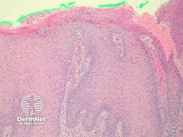
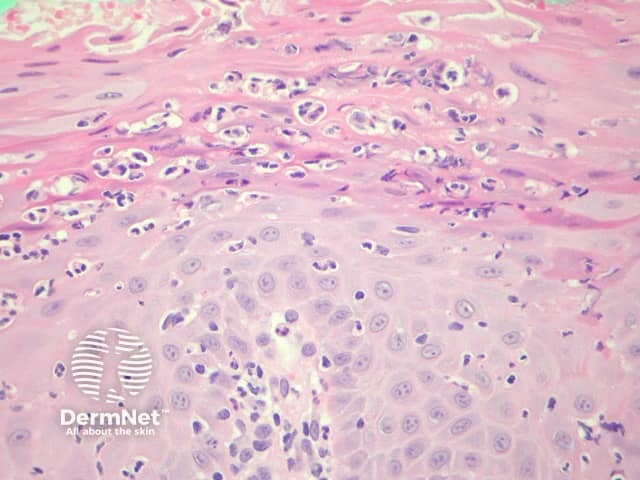
In candidiasis, sections show predominantly spongiotic changes in the epidermis with irregular acanthosis, mild spongiosis and inflammatory changes (figure 1). In the superficial epidermis, the characteristic feature is the presence of neutrophils in the stratum corneum and upper layers of the epidermis. The neutrophils may form small collections (spongiform postulation) which resembles impetigo or psoriasis (figure 2).
Special stains for candidiasis
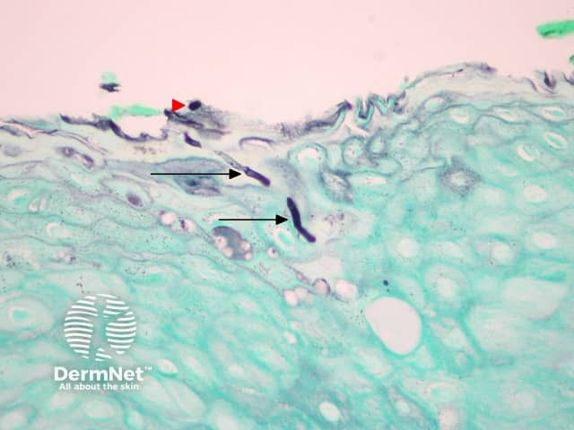
Special stains to demonstrate the yeast forms are essential in candidiasis.
- Gomori methenamine silver stain (GMS). The stain highlights the pseudohyphal or hyphal forms (figure 3, arrows) penetrating into the keratinized epithelium. In addition, there are yeast-like forms in the superficial stratum corneum (figure 3, red arrowhead).
- PAS stain can also be used to highlight the organisms.
Differential diagnosis of candidiasis pathology
Pustular psoriasis, subcorneal pustolosis, acute generalized subcorneal pustulosis – Spongiform postulation can also be seen in these conditions. Special stains should be carefully examined in cases of psoriasis to exclude a fungal aetiology.
Impetigo – Spongiform postulation is a hallmark of impetigo. Gram stain may highlight bacterial colonies in impetigo which GMS and PAS stains will fail to reveal fungal forms.
Dermatophytosis – Spongiform postulation is a hallmark of tinea cruris and corporis. Special stains show septate hyphae without the budding yeasts of candida. Distinction can sometimes be difficult. Candida often penetrates the keratinized epithelium (figure 3) as opposed to dermatophytosis which usually involves just the stratum corneum.
References
- Weedon’s Skin Pathology (Third edition, 2010). David Weedon
- Pathology of the Skin (Fourth edition, 2012). McKee PH, J. Calonje JE, Granter SR
On DermNet