Main menu
Common skin conditions

NEWS
Join DermNet PRO
Read more
Quick links
Lesions (benign) Diagnosis and testing
Author: Dr Ben Tallon, Dermatologist/Dermatopathologist, Tauranga, New Zealand, 2012.
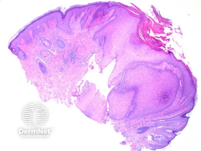
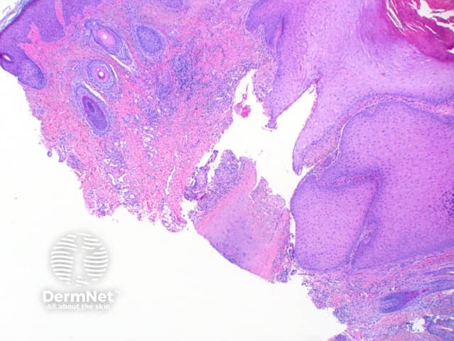
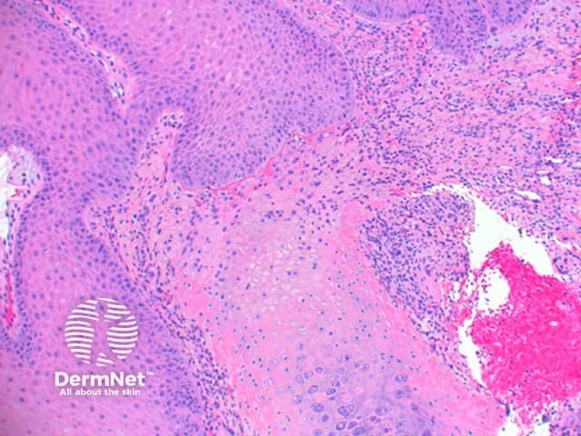
Scanning power view of chondrodermatitis nodularis helicis shows a wedge-shaped dermal alteration extending through to the superficial cartilage (Figure 1). The epidermis may show a focal zone of orthokeratosis and scale crust overlying moderate epidermal hyperplasia (Figure 2). Frequently there will be superficial epidermal ulceration. There is a zone of eosinophilic fibrinoid material overlying the affected area of cartilage, with the blurring of the superficial cartilage and altered staining (Figure 3). There may be adjacent perichondrial thickening.

Figure 1

Figure 2

Figure 3
Pseudocyst of the auricle: while there may also be hyalinised degeneration above the affected collagen, a non-epithelial lined cystic space forms within the collagen in pseudocyst of the auricle.