Conjunctivitis is inflammation of the conjunctiva of the eye.
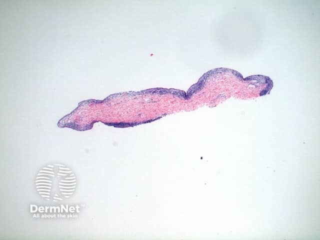
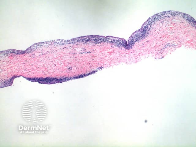
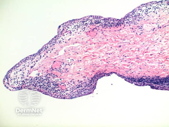
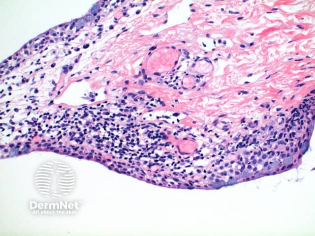
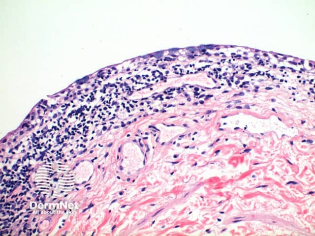
The low power view of the histology of conjunctivitis shows a delicate specimen with a mild spongiotic reaction pattern (figure 1). The conjunctival epithelium is comprised of a stratified squamous epithelium with numerous interspersed goblet cells containing mucin (figures 2-5). Closer inspection reveals spongiosis of the epithelium with lymphocytic exocytosis (figures 3, 4). The subepithelial tissue contains telangiectatic vessels and a mild superficial perivascular lymphocytic infiltrate with occasional to numerous plasma cells (figure 5).
Allergic conjunctivitis: the presence of numerous eosinophils is the discriminating feature.
Infectious conjunctivitis: typically the presence of numerous neutrophils should raise concern for a bact erial conjunctivitis. On histology this cannot be excluded and culture should be recommended if there is any clinical concern. Chlamydial conjunctivitis (among others) may have identical histologic features to chronic conjunctivitis.
Chronic conjunctivitis: the epithelium is hyperplastic and there are increased numbers of plasma cells.
Ligneous (pseudomembranous) conjunctivitis: In this variant there are additional deposits of pink amorphous material within the subepithelial tissue, which may mimic amyloid deposits, contiguous with a chronic inflammatory cell infiltrate. Clinically there is pseudomembrane formation, which may affect other mucosal surfaces. The histological pseudomembrane typically separates from the underlying epithelium without bleeding.
Giant papillary conjunctivitis: the low power shows a papillomatous projection of conjunctival epithelium which otherwise shows features of chronic conjunctivitis.

