Human gnathostomiasis presents as larva migrans profundus, and is caused by ingesting gnathostoma larvae. It is characterised by cutaneous migratory swellings. The larvae are ingested via raw fish in warmer climates in South America and parts of Asia.
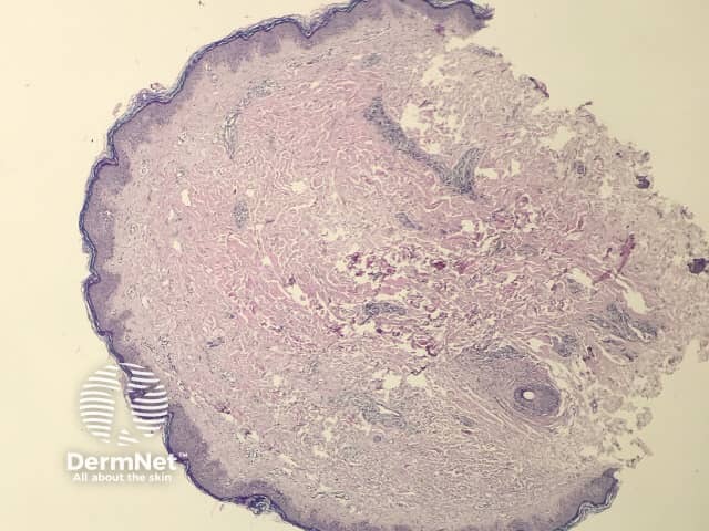
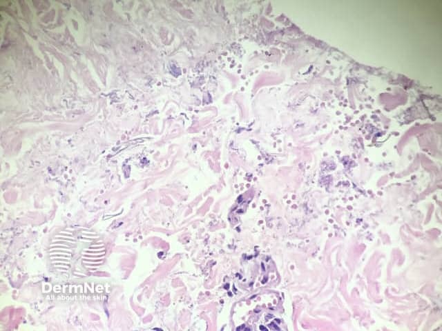
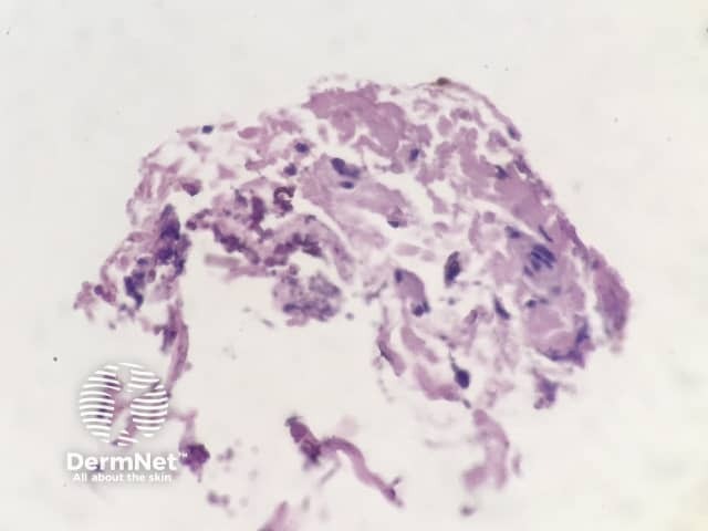
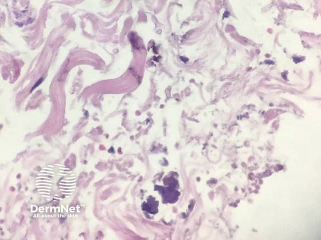
Histopathology of gnathostomiasis shows a diffuse inflammatory cell infiltration in the dermis with eosinophils and denatured collagen fragments (figures 1–4). Parasite parts may be seen.
None are generally useful.
The diagnosis of gnathostomiasis is difficult without clinical correlation. The reaction pattern is identical to that seen in an exaggerated bite reaction or reaction to a variety of parasites. Identification of parasitic parts can also be difficult to define as gnathostomiasis in routine haematoxylin and eosin sections.
The enzyme‐labelled antibody method for a serum‐specific antibody against gnathostoma species shows positive results.
Yamanaka M, Kusutani N, Sowa-Osako J, et al. Gnathostomiasis caused by ingestion of raw Oncorhynchus masou ishikawae roe. J Dermatol 2017; 44: e208–9. DOI: 10.1111/1346-8138.13893. PubMed

