

Infantile digital fibromatosis pathology — extra information
Introduction Histology Special studies Differential diagnoses
Introduction
Infantile digital fibroma, also called inclusion body fibromatosis or Reye tumour, is a benign proliferation of myofibroblasts.
Histology of infantile digital fibromatosis
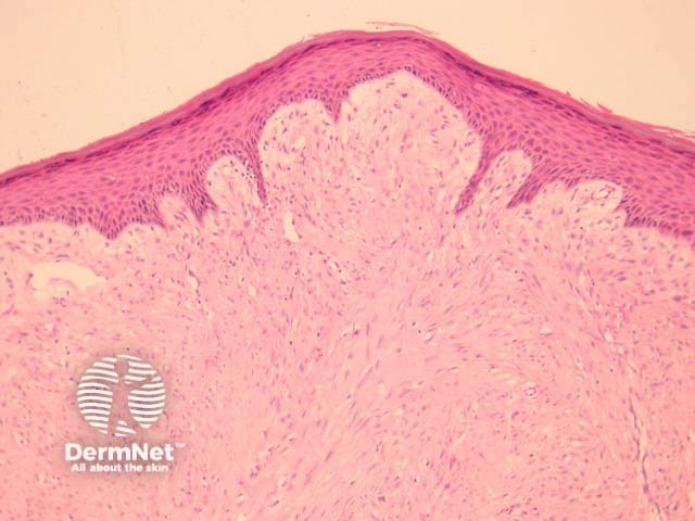
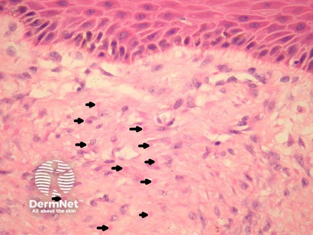
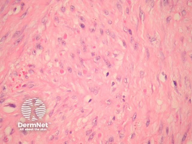
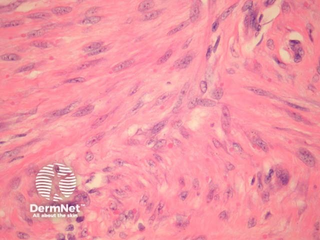
In infantile digital fibromatosis, sections show an intradermal unencapsulated tumour composed of spindle-shaped cells arranged in whorls or interdigitating sheets (figure 1). These myofibroblasts contain 3–10 μm inclusion bodies, which are round or ovoid and granular (figures 2-4, arrows are used to highlight some of the inclusion bodies in figure 2).
Earlier lesions of infantile digital fibromatosis are more inflammatory; more developed lesions display more fibroplasia and inclusion bodies.
Special studies for infantile digital fibromatosis
The inclusion bodies stain pink with H&E (figures 2-4). The bodies are positive with immunohistochemical stains for actin and vimentin.
Differential diagnosis of infantile digital fibromatosis pathology
Fibromatosis – Identification of the characteristic inclusion bodies distinguish infantile digital fibromatosis from dermal fibromatosis and hypertrophic dermal scars
References
- Grenier N, Liang C, Capaldi L, Ney A, Lapidus C, Schappell D, Katarincic J, Robinson-Bostom L. A range of histologic findings in infantile digital fibromatosis. Pediatr Dermatol. 2008 Jan-Feb;25(1):72–5. PubMed
On DermNet