Lipoid proteinosis is a rare autosomal recessive disease caused by mutations in the extracellular matrix protein 1 gene (ECM1).
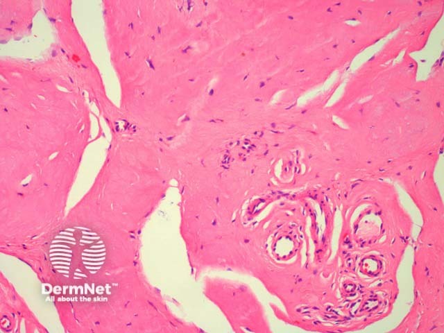
In lipoid proteinosis, sections show deposition of an eosinophilic homogenous material in the dermis (figures 1, 2). The overlying epidermis may be papillomatous (figure 1). The material surrounds blood vessels and adnexa (figure 2).
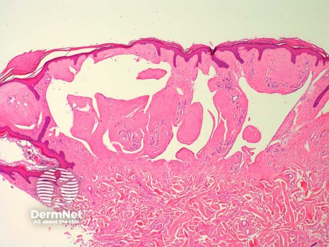
Sweat gland hyalinisation and hyperkeratosis, and epidermal acanthosis may also be seen.
The eosinophilic material is strongly positive with periodic acid-Schif staining (PAS) and weakly positive or negative for congo red and thioflavine-T.
Colloid milium and amyloidosis – both conditions show accumulation of a similar eosinophilic material. These should both be congo red positive. Clinical history can be helpful to exclude colloid milium.
Immunohistochemical studies for keratin and other amyloid-associated proteins can be helpful if amyloidosis is suspected. immunohistochemical staining may show absence or attenuation of ECM1; labelling with an anti-collagen antibody shows types IV and V collagen accumulation around blood vessels.
Electron microscopy reveals reduplication and disruption of the basement membranes at the DEJ perivascularly. Electron microscopy of early vesicular lesions may also show ‘free-floating’ desmosomes within the intercellular space between keratinocytes.

