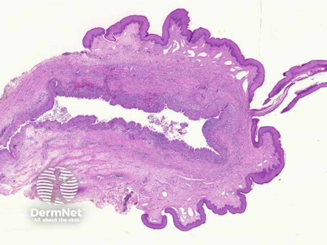
Median raphe cysts are developmental and usually arise on the ventral surface of the penis of young men. They are thought to represent either abnormal closing of the median raphe or abnormal separation of the uretheral epithelium during development.
The median raphe cyst is in the dermis and does not drain into the overlying epidermis (figure 1).
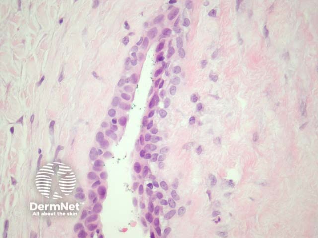
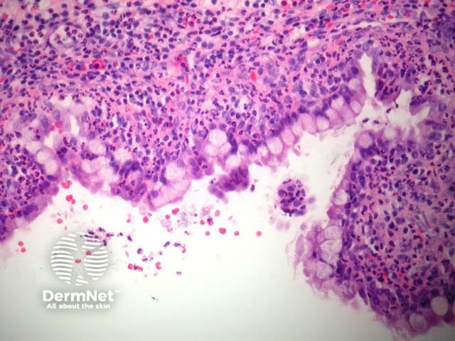
The lining of the cyst is usually a pseudostratified columnar epithelium (figure 2). Some cases may have mucinous glandular epithelium (figure 3) or a mixture of mucinous, transitional, and squamous epithelia. Cilia are usually not seen. At times, the lining epithelium may be entirely denuded.
None are needed.
The anatomic location of a median raphe cyst usually allows accurate distinction from other developmental cysts such as bronchogenic cyst and omphalomesenteric cyst. Bartholin duct cysts show a similar pseudostratified epithelium.

