

Mucinous carcinoma pathology — extra information
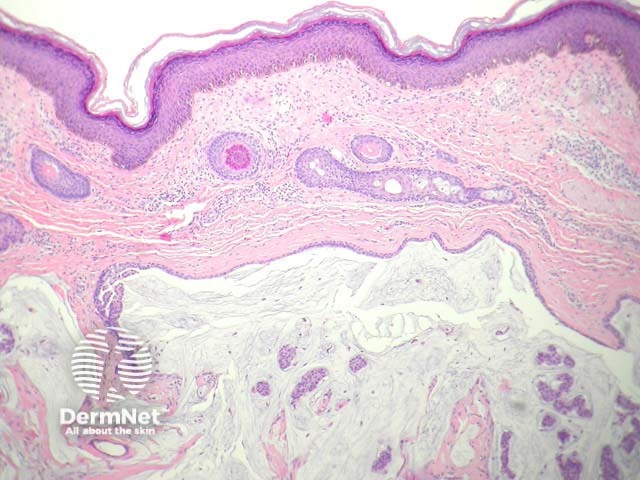
Primary cutaneous mucinous carcinoma is a distinctive tumour that has been variously described as apocrine and eccrine. The most frequent described presentation has been a slow-growing dermal mass arising on the head and neck of older adults.
Histology of mucinous carcinoma
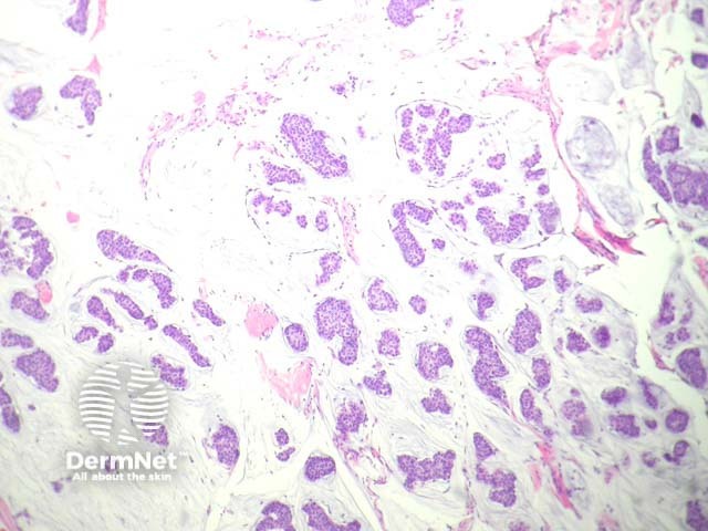
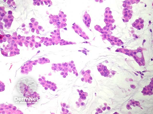
In mucinous carcinoma, there is a dermal mass composed of glands and solid nests of epithelial cells which appear to be floating in copious mucin (figures 1–3). The cells are enlarged, show mild pleomorphism and often have an eosinophilic cytoplasm (figure 3).
Special studies for mucinous carcinoma
Immunohistochemical studies reveal the tumour is positive with CK7, and may express ER (oestrogen receptor) and PR (progesterone receptor). p63 and CK5/6 may highlight a population of myoepithelial cells and help to distinguish this tumour from a cutaneous metastasis from another site.
Differential diagnosis of mucinous carcinoma
Cutaneous metastasis from an internal malignancy – Colloid carcinoma of the breast and other mucinous carcinomas of the ovary, pancreas and other sites may be morphologically identical. Clinical correlation can be helpful. Immunohistochemical studies with CK5/6, and p63 have been advocated as being useful – any positivity for these antibodies favour a primary cutaneous tumour rather than metastasis from another site. Negativity with CK7 should raise the possibility of metastasis from another site.
Endocrine mucin-producing sweat gland carcinoma – This tumour is thought by some authors to represent precursors to mucinous carcinoma. They are composed of solid nests of cells which exhibit some neuroendocrine differentiation. These tumours arise almost exclusively in the skin surrounding the eye
References
- Levy G, Finkelstein A, McNiff JM. Immunohistochemical techniques to compare primary vs. metastatic mucinous carcinoma of the skin. J Cutan Pathol. 2010 Apr;37(4):411–5. PubMed
On DermNet