Nodular fasciitis presents as a rapidly growing soft tissue mass which may follow a history of trauma.
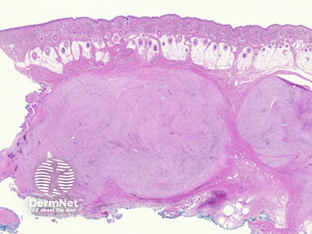
Low power examination of nodular fasciitis shows a well-circumscribed discrete mass in the subcutaneous adipose tissue (figure 1). Dermal and intravascular forms have also been described.
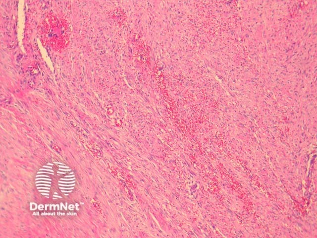
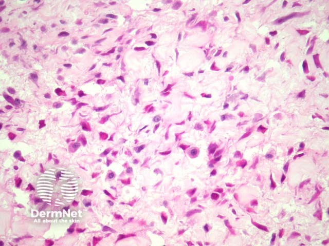
The tumour is composed of haphazardly arranged mass of spindled and plump cells which is often compared to tissue culture. The cells are set in a fibromyxoid stroma. Some typical mitoses are commonly seen. There may be numerous red blood cells (figure 2), and chronic inflammatory cells. Proliferative fasciitis is a well recognized variant and is composed of epithelioid cells which resemble ganglion cells (figure 3).
The myofibroblasts are positive with smooth muscle actin, calponin. Desmin and S100 are negative
Sarcoma – Atypical mitoses, tumour necrosis and frank nuclear atypia are unusual in nodular fasciitis and suggest a malignant process. Low grade sarcomas (such as low grade fibromyxoid tumour) may cause diagnostic difficulty but generally do not exhibit the tissue culture-like disorganized array of myofibroblasts.

