Diagnosis and testing Connective tissue diseases
Palmar fibromatosis and plantar fibromatosis represent essentially the same process effecting hands and feet respectively.
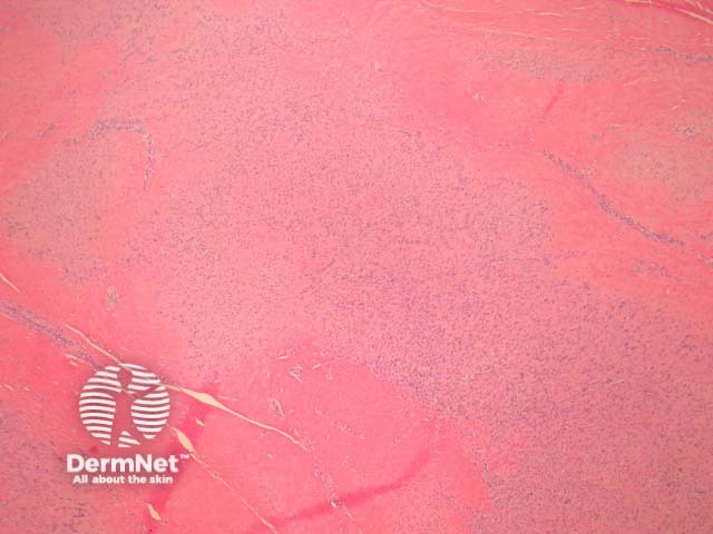
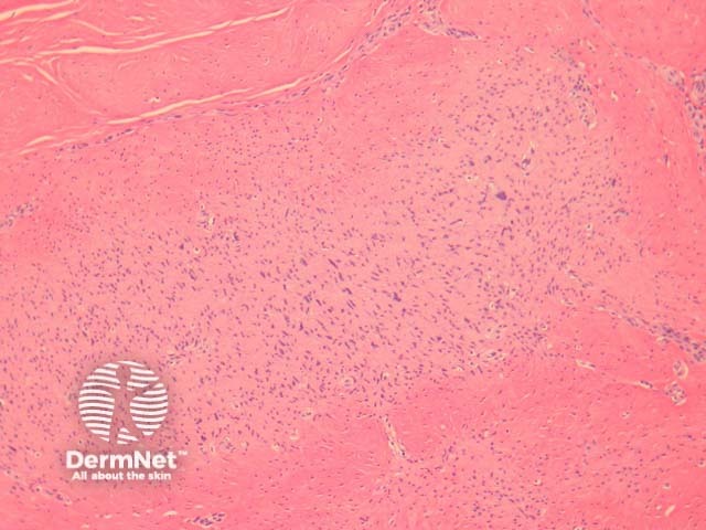
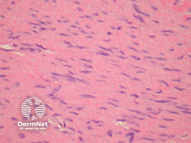
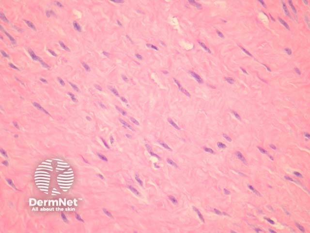
In palmoplantar fibromatosis, sections show a dense fibrocellular tissue with mature collagen and nodular collections of myofibrocytes in various stages of maturation. There may be some nuclear atypia and mitotic activity but the cells lack prominent atypical features or abnormal mitotic figures. The proliferating cells are plump, with elongated vesicular nuclei and small nucleoli (figures 1-4).
Unusual findings include focal bone formation and giant cells.
None are generally needed. The proliferating cells are myofibroblasts rather than fibroblasts and thus will stain with smooth muscle markers.
Clinical location may distinguish palmoplantar fibromatosis from other fibromatoses such as knuckle pads, desmoid tumours, and penile fibromatosis (Peyronie disease). Interestingly, deep fibromatoses are genetically distinct and harbour mutations in CTNNB1 which encodes beta-catenin.
Infantile digital fibromatosis – This juvenile form of fibromatosis is distinguished by identification of the characteristic inclusion bodies.

