DermNet provides Google Translate, a free machine translation service. Note that this may not provide an exact translation in all languages
Home » Topics A–Z » Perpendicular white lines dermoscopy
Author: Naomi Ashman, Dermoscopist, Torbay Skin, Auckland, New Zealand. DermNet NZ Editor in Chief Adjunct A/Prof Amanda Oakley, Dermatologist, Hamilton, New Zealand. November 2019.
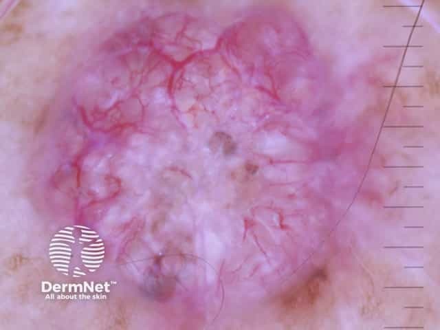
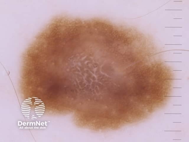
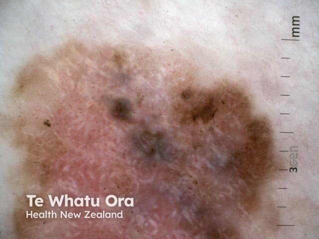
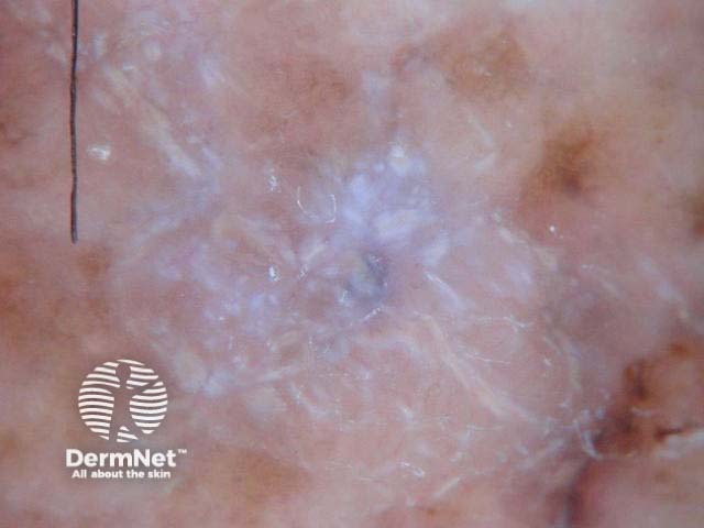
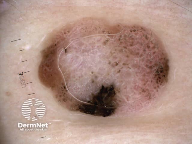
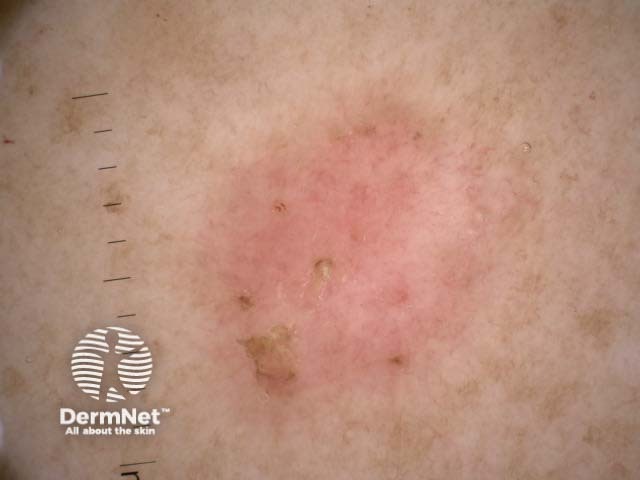
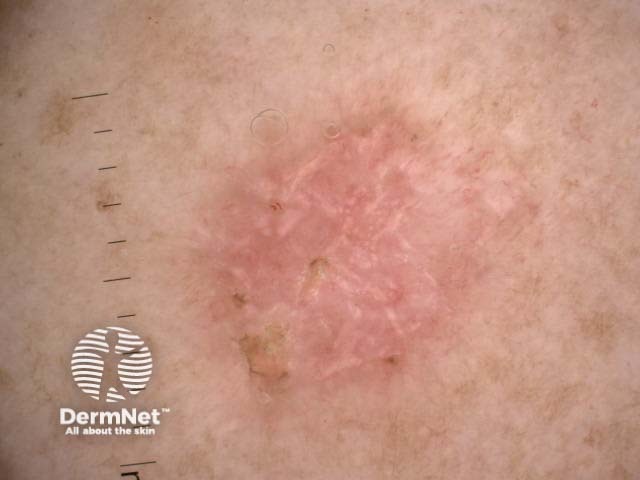
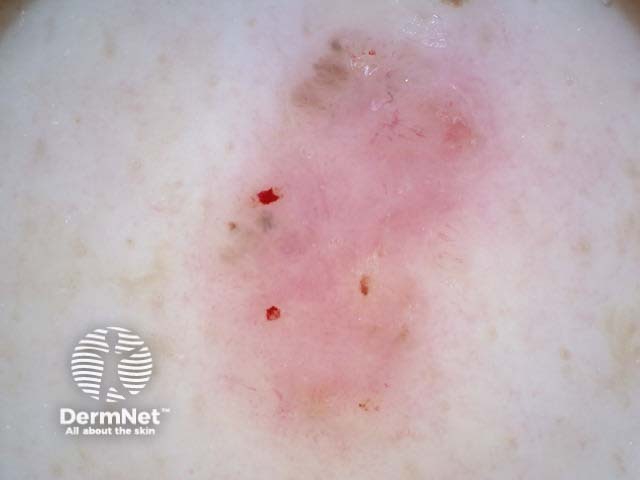
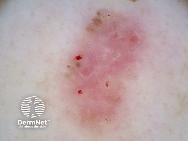
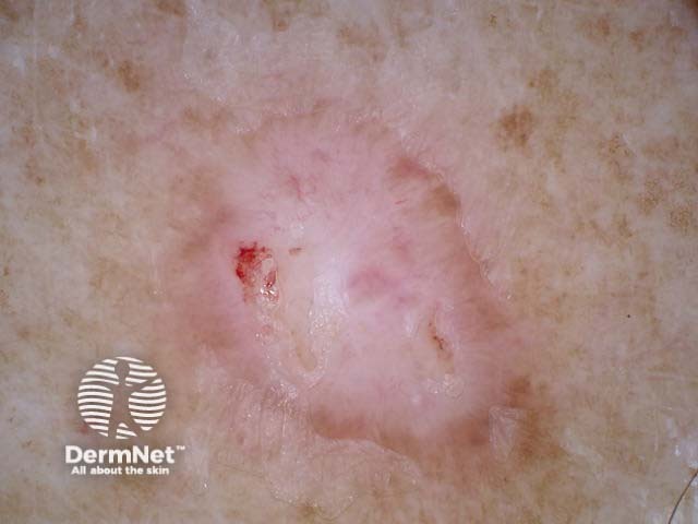
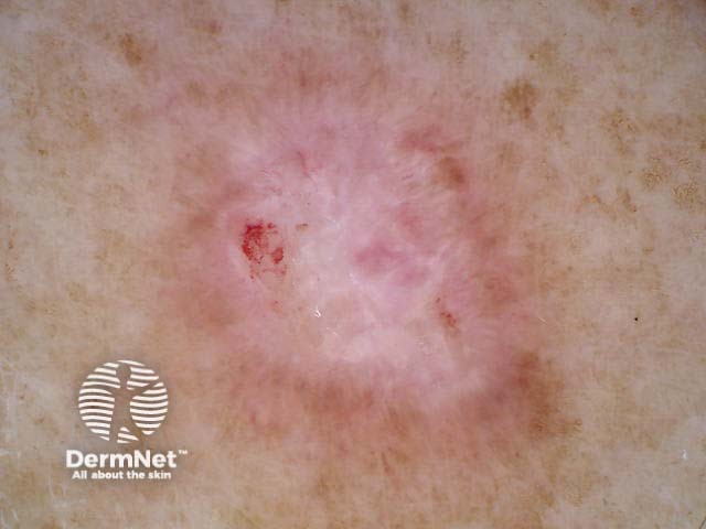
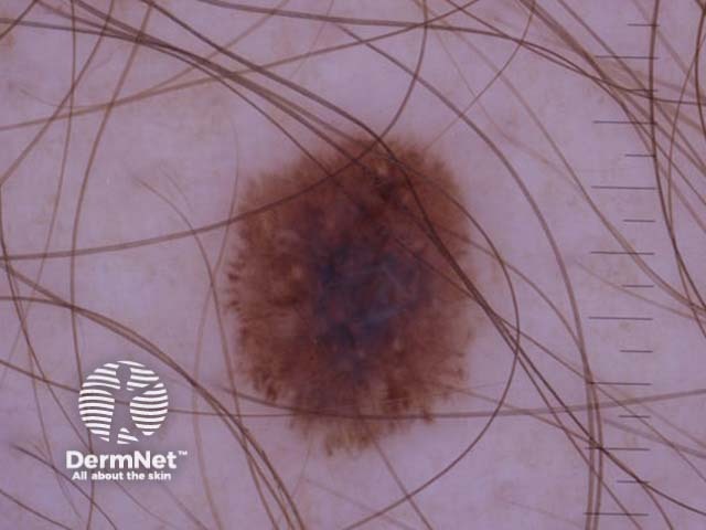
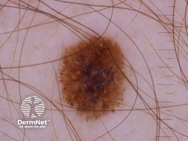
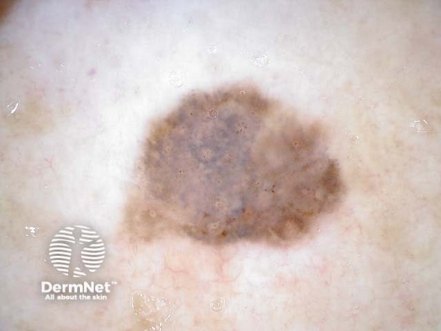
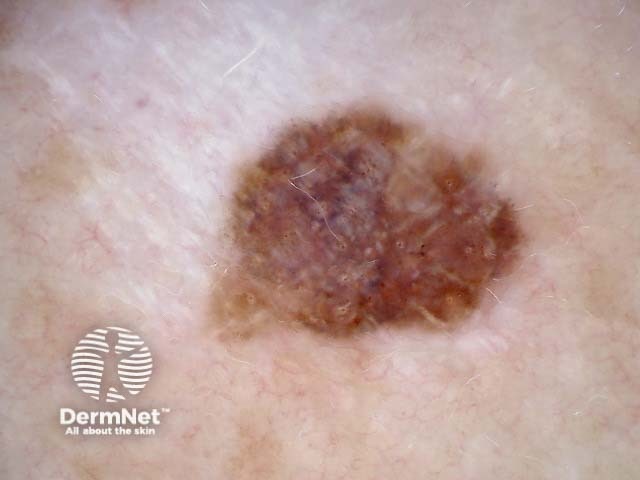
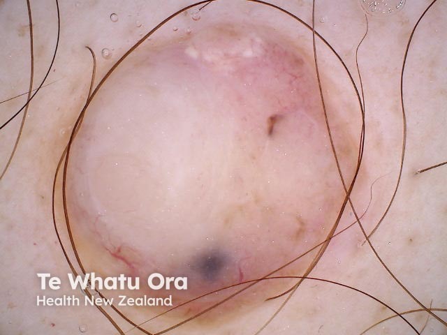
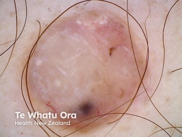
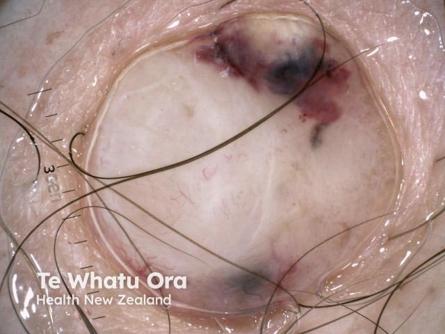
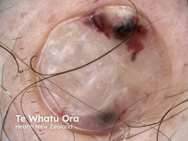
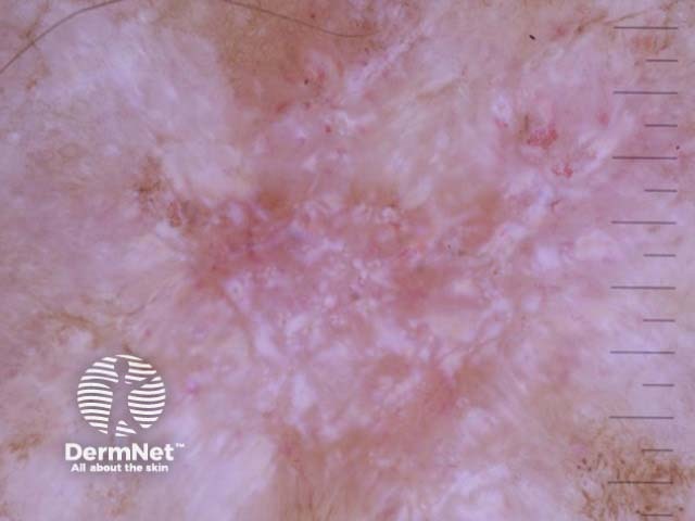
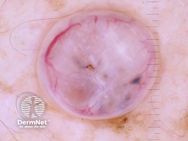
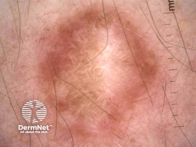
In dermoscopy, perpendicular white lines are short discrete white lines oriented parallel and orthogonal (perpendicular) to each other and seen only under polarised light [1]. They are also known as polarising white lines, short white lines, shiny white lines, shiny white streaks, chrysalis, chrysalids, and crystalline structures. Perpendicular white lines are a clue to a specific diagnosis including basal cell carcinoma (BCC) and some melanomas [2].
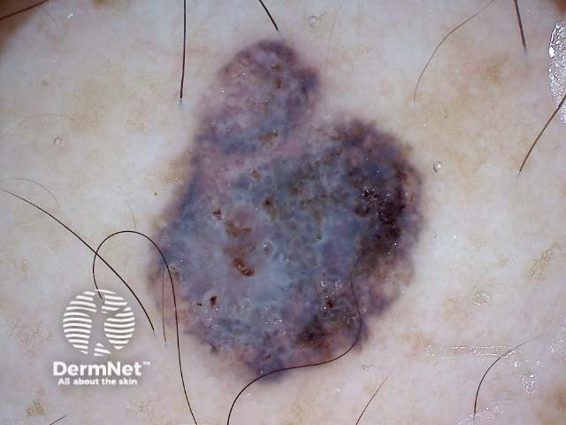
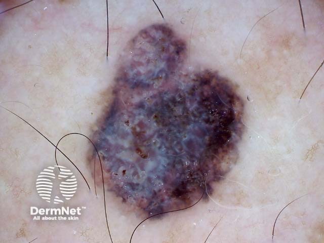
Perpendicular white lines are only seen under polarised light. They appear as short, shiny white lines and move as the dermoscopy lens is moved at different angles over the lesion.
Irregular pinkish lesion with erosions, short red vessels, erosions, polarised white structures Superficial basal cell carcinoma dermoscopy Superficial basal cell carcinoma dermoscopy Ill-defined borders, skin coloured stroma, branched red lines, prominent polarised white lines Pink to purple stroma, prominent irregular vessels, greyish clods, prominent polarised white structures Central ulceration, ill-defined pinkish stroma, few short vessels, solitary bluish structure, and polarised white linesPerpendicular white lines in BCCs
Perpendicular white lines in melanoma
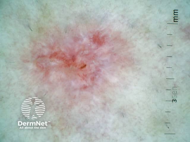
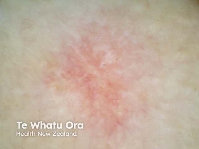
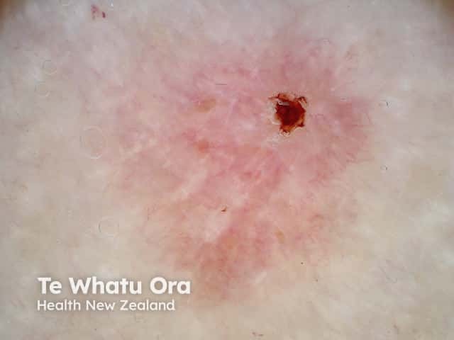
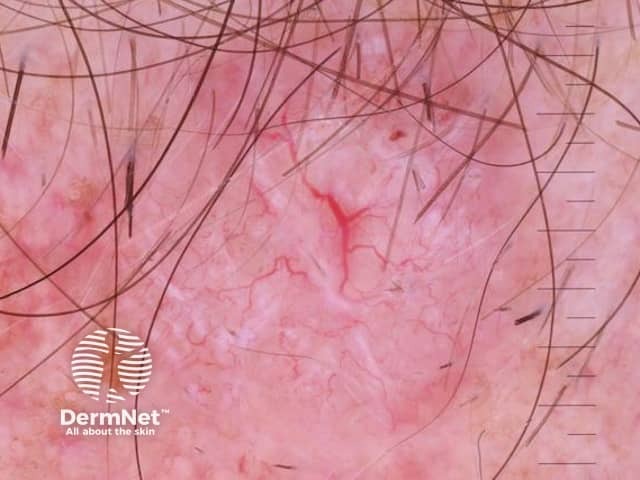
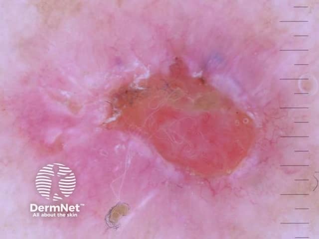
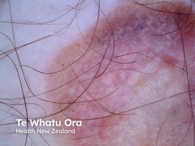
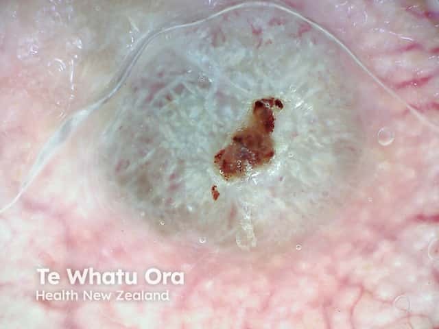
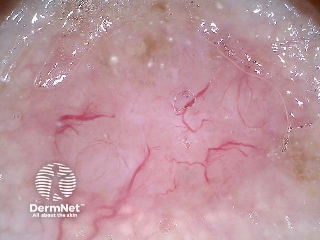
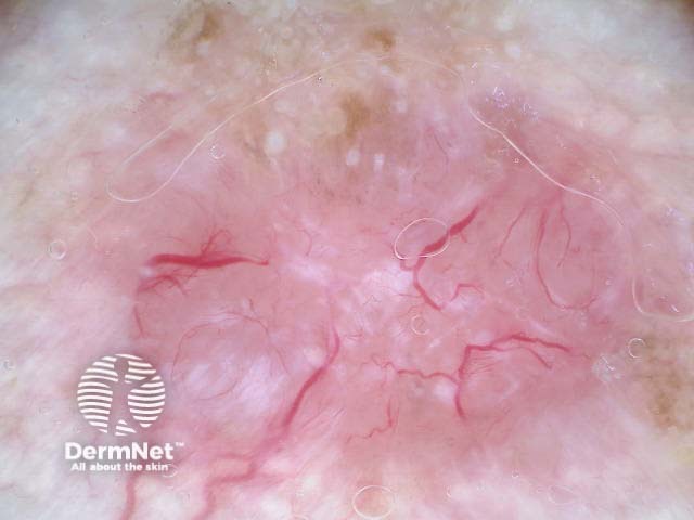
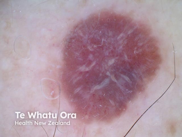
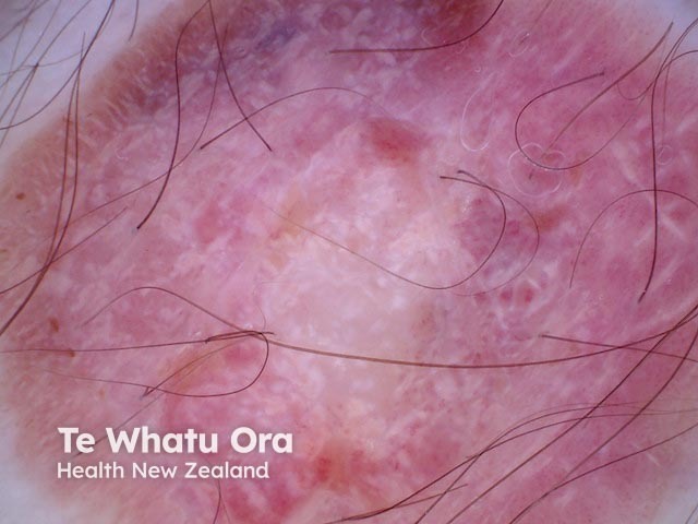
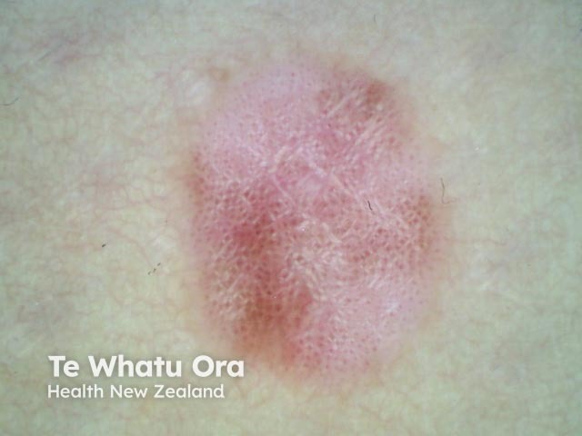
The following pairs of images demonstrate the differences seen in dermoscopy of perpendicular white lines under polarised and nonpolarised light.
Polarised and nonpolarised light in BCC
Perpendicular white lines are absent in a melanoma in situ when viewed under nonpolarised light dermoscopy Perpendicular white lines are visible under polarised light dermoscopy of a melanoma in situ Melanoma in situ, nonpolarised dermoscopy view Melanoma in situ, polarised dermoscopy view Nonpolarised dermoscopy view of nodular melanoma Polarised dermoscopy view of nodular melanoma Nonpolarised dermoscopy view of nodular melanoma, 7.2 mm thick Polarised dermoscopic view of nodular melanoma, 7.2 mm thick Nonpolarised dermoscopy of melanoma, 0.8 mm thick Polarised dermoscopy of melanoma, 0.8 mm thickPolarised and nonpolarised light in melanoma
Perpendicular white lines can be seen in the following lesions:
Red irregular patch with short linear vessels, some red dots, and polarised white perpendicular lines Large irregular pink plaque with scattered light brown concentric and short linear structures with prominent polarised white structures Superficial basal cell carcinoma dermoscopy Irregular lesion with pink background, superficial erosions, peripheral pigmented structures, peppering, polarised white lines Nodular basal cell carcinoma dermoscopy Polarised dermoscopy of a nodular basal cell carcinoma presenting as an exophytic polyp Invasive melanoma dermoscopy, Breslow 0.4mm within a melanoma in situ, with associated naevus present Amelanotic melanoma dermoscopy Amelanotic melanoma dermoscopy Spitz naevus dermoscopy Dermatofibroma Dermatofibroma dermoscopyPerpendicular white lines in a variety of lesions
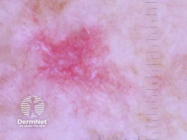
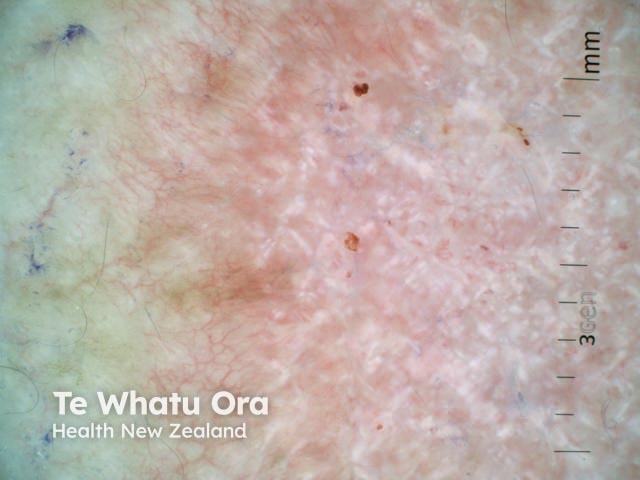
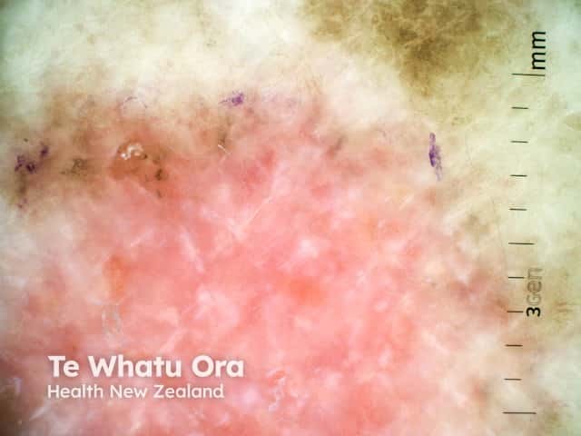
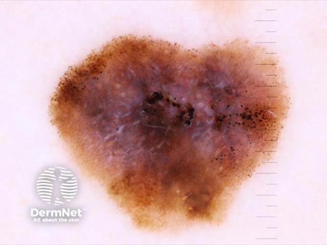
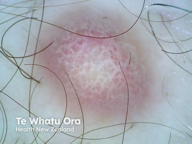
Perpendicular white lines are thought to correlate histopathologically with altered collagen in the dermis (fibrosis). The birefringent properties of collagen bundles cause rapid randomisation of polarised light. This is the reason collagen appears bright white and is more conspicuous under polarised dermoscopy [3].
They also correlate with dermal invasion in cases of melanoma [4]. However, as our illustrations show, they may also be seen in melanoma in situ.
See smartphone apps to check your skin.
[Sponsored content]
© 2021 DermNet New Zealand Trust.
DermNet NZ does not provide an online consultation service. If you have any concerns with your skin or its treatment, see a dermatologist for advice.