

Pigmented follicular cyst pathology — extra information
Categories:
ICD-10:
ICD-11:
SNOMED CT:
ADVERTISEMENT
This benign lesion is grouped within the cutaneous cysts
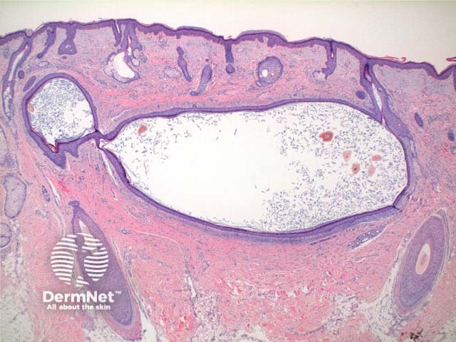
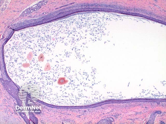
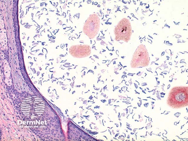
Histology of pigmented follicular cyst
Scanning power view demonstrates a unilocular cystic structure within the dermis (Figure 1). Higher power reveals a thin epithelial lining with a retained granular layer (Figures 2 and 3). Within the cyst cavity are loose keratin fragments and numerous pigmented terminal hair shafts (Figures 2 and 3).
Differential diagnosis of pigmented follicular cyst
Vellous hair cyst: In this lesion the cyst cavity contains small vellous hairs lacking pigmentation.
Epidermal inclusion cyst: Hair fragments are absent.
References
- Skin Pathology (2nd edition, 2002). Weedon D
- Pathology of the Skin (3rd edition, 2005). McKee PH, J. Calonje JE, Granter SR
On DermNet
Books about skin diseases
ADVERTISEMENT
Other recommended articles
ADVERTISEMENT