

Scabies pathology — extra information
Histology of scabies
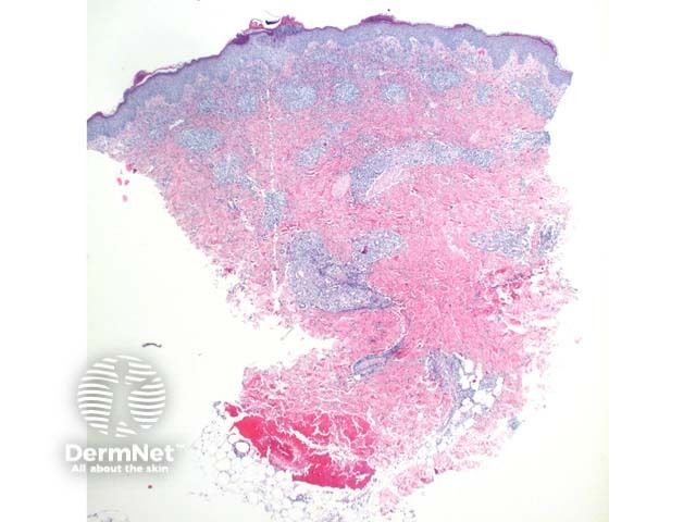
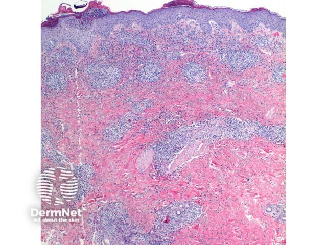
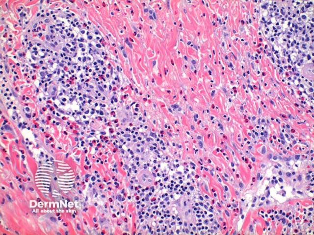
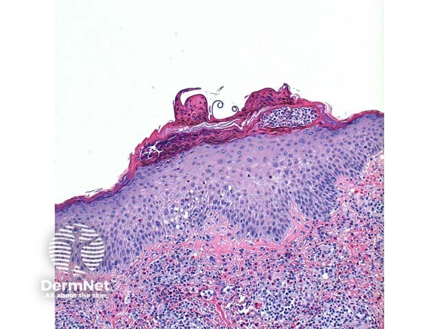
Scanning power view of scabies shows a pattern of an epidermal and wedge shaped dermal inflammatory process (Figure 1). The epidermis may show significant scale crust comprised of serous exudate, neutrophils, and eosinophils (Figure 2). There may be focal ulceration or erosion secondary to excoriation. The inflammatory infiltrate may show a wedge shaped or diffuse superficial and deep perivascular and interstitial pattern. Lymphocytes with numerous eosinophils are the rule with scattered superficial neutrophils seen in excoriated or impetiginised cases. Deep interstitial eosinophils are an important clue to an arthropod bite reaction (Figure 3).
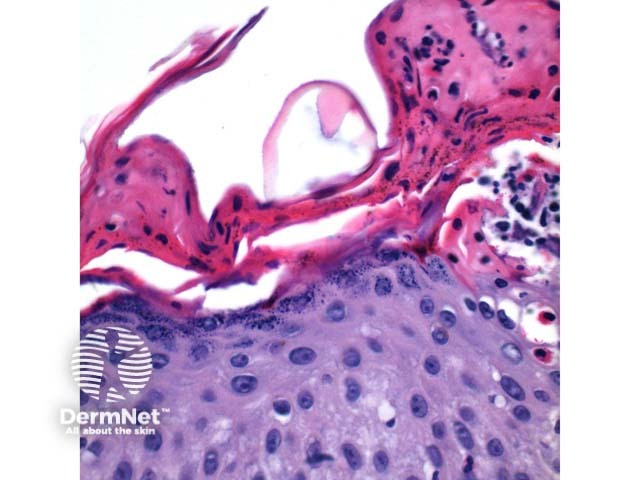
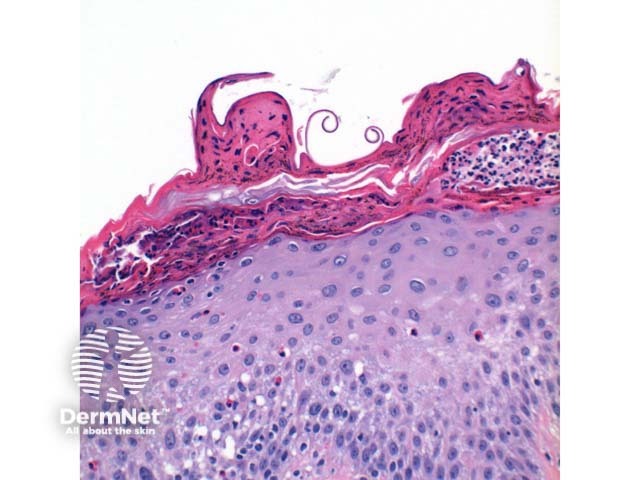
The presence of scabies parts evident as solid pink eosinophilic fragments is diagnostic, representing the chitinous exoskeleton of the Sarcoptes scabiei mite (Figure 4). Pink pigtails can also be seen occasionally within the stratum corneum, and are thought to represent remnants of the egg shell (Figures 5 and 6).
Histological variants of scabies
In crusted (Norwegian) scabies, the epidermis shows psoriasiform hyperplasia with marked hyperkeratosis.
Differential diagnosis of scabies
Differential diagnosis of scabies include the following conditions.
Bullous pemphigoid
The urticarial phase of bullous pemphigoid can show similar features to scabies, though typically the infiltrate is superficial with lining up of the eosinophils along the basal layer of the epidermis and mild vacuolar change evident. It is important to remember that positive linear basement membrane staining on direct immunofluorescence testing has been reported in scabies infestation.
Allergic contact dermatitis
The presence of spongiosis with vesiculation, while favouring allergic contact dermatitis, is also seen in scabies and so cannot be relied upon as a differentiating factor. A superficial infiltrate with numerous eosinophils within the epidermis may serve as a clue.
Fibreglass dermatitis
Fibreglass can be identified within the stratum corneum. It largely causes an irritant dermatitis with eosinophils a less dominant feature in fibreglass dermatitis.
References
- Skin Pathology (3rd edition, 2002). Weedon D
- Pathology of the Skin (3rd edition, 2005). McKee PH, J. Calonje JE, Granter SR
- Pathologic findings in human scabies. Arch Dermatol 1977 Mar; 113: 320–4. PubMed
On DermNet
- Crusted scabies pathology
- Scabies
- Crusted scabies
- Bullous pemphigoid
- Allergic contact dermatitis
- Fibreglass dermatitis
- Dermatopathology glossary
- Dermatopathology index