DermNet provides Google Translate, a free machine translation service. Note that this may not provide an exact translation in all languages

Translate

Sclerema neonatorum pathology

Author: Assoc Prof Patrick Emanuel, Dermatopathologist, Auckland, New Zealand. January 2015.


toc-icon
Table of contents
arrow-right-small

Sclerema neonatorum usually affects gravely ill, preterm neonates in the first week of life. It is characterised by hardening of the skin that gets bound down to the underlying structures.

Histology of sclerema neonatorum

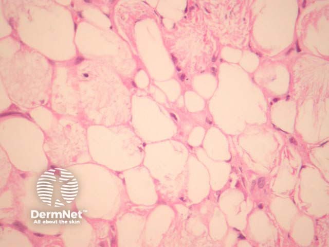
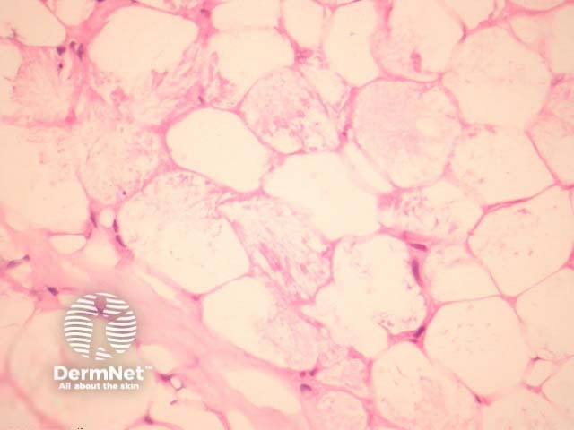
In sclerema neonatorum, needle-shaped crystals are arranged radially in adipocytes. There is little or no associated inflammatory infiltrate and no fat necrosis (figures 1, 2).

Sclerema neonatorum pathology

Special studies for sclerema neonatorum

None are generally needed.

Differential diagnosis of sclerema neonatorum

Poststeroid panniculitis and subcutaneous fat necrosis of the newborn: These show identical crystals but there is associated inflammatory and foreign body response.


Bibliography

  • Weedon's Skin Pathology (Third edition, 2010). David Weedon
  • Zeb A, Darmstadt GL. J Perinatol. Sclerema neonatorum: a review of nomenclature, clinical presentation, histological features, differential diagnoses and management. 2008 Jul;28(7):453–60. PubMed

On DermNet

Books about skin diseases


Related information

Sign up to the newsletter