DermNet provides Google Translate, a free machine translation service. Note that this may not provide an exact translation in all languages

Translate

Splenosis pathology

Author: Adjunct A/Prof Patrick Emanuel, Dermatopathologist, Clínica Ricardo Palma, Lima, Peru. DermNet NZ Editor in Chief: Adjunct A/Prof Amanda Oakley. Copy edited by Gus Mitchell. September 2018.


toc-icon
Table of contents
arrow-right-small

Introduction

Splenosis may present in the skin following splenic trauma and subsequent spread of benign splenic tissue into surrounding structures.

Histology of splenosis

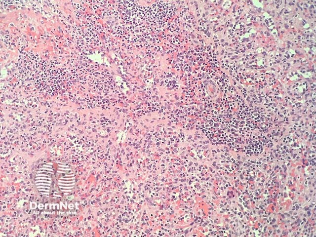
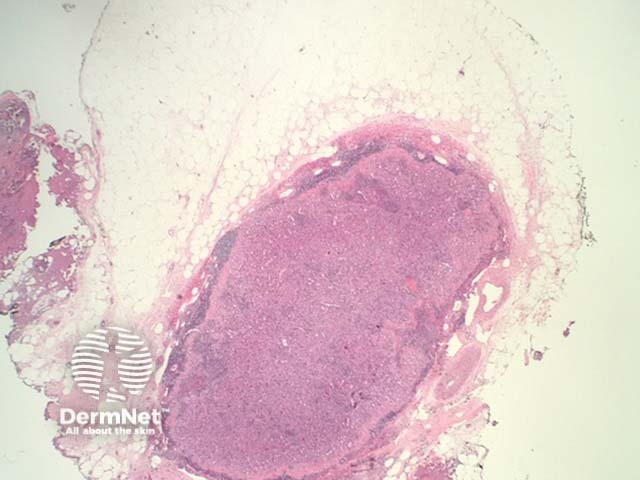
In splenosis, the histopathology shows normal splenic tissue, usually located in the subcutis (figures 1, 2). Spleen can be recognised by the presence of red pulp (composed of venous sinusoids allowing passing of red blood cells) and white pulp (composed of sheaths of lymphoid cells around arteries).

Splenosis pathology

Special studies for splenosis

None are generally needed.

Differential diagnosis for splenosis

Other diagnoses to be considered include a dermal lymph node: this lacks the components seen in splenic tissue (red and white pulp).


References

  • Gupta K, Ahluwalia A, Jain T, Saggar K. Abdominal splenosis mimicking peritoneal deposits- A case report. The Pan African Medical Journal 2014; 17: 269. doi:10.11604/pamj.2014.17.269.3413. PubMed Central

On DermNet

Other websites

Books about skin diseases


Related information

Sign up to the newsletter