Created 2008.
Develop skills in examining the nails and describing:
This section provides a glossary of terms used to describe abnormal fingernails and toenails. Proper use of language is necessary for diagnosis and to communicate with other health professionals.
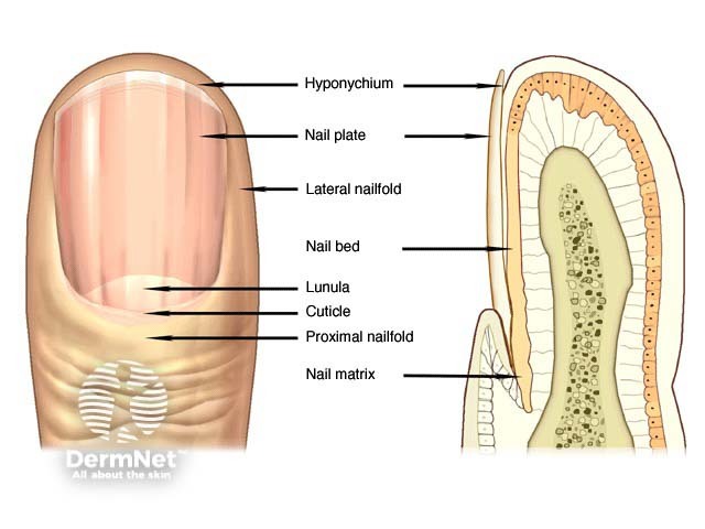
Nails are a specialised form of stratum corneum and are made predominantly of keratin. Their primary functions are for protection, scratching, and picking up small objects. When looking at the nails carefully inspect the nail plate and surrounding skin.
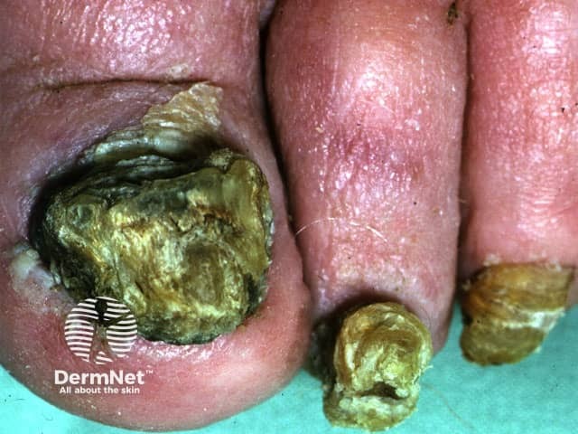
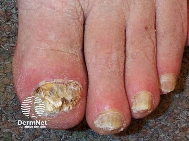
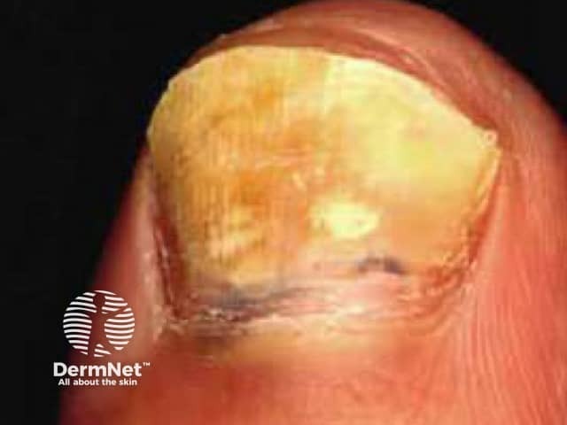
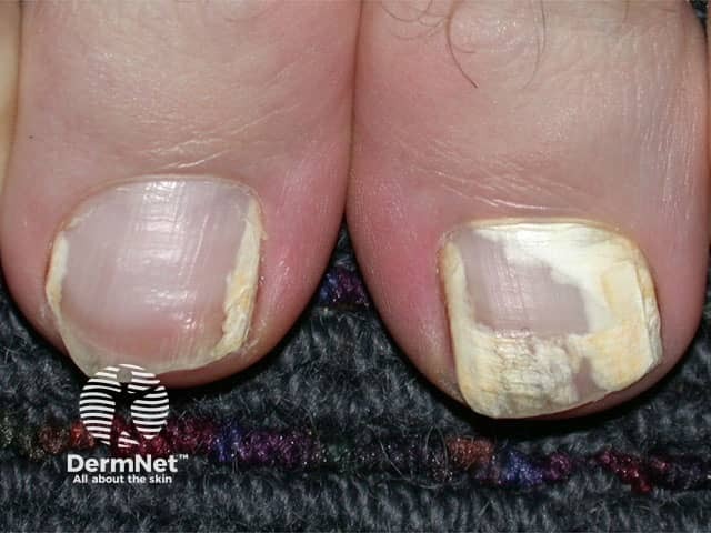
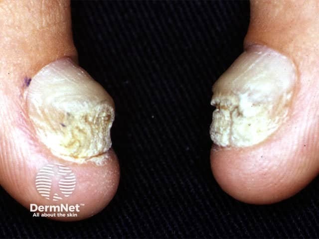
If the patient presents with a nail problem, it is important to ask about skin disease elsewhere and examine them generally. Fungal nail disease (onychomycosis) is nearly always associated with fungal skin disease (check feet, hands, groin). Nail changes may be the first sign of psoriasis (check scalp, elbows, knees, flexures), lichen planus (check oral mucosa, lower back, scalp, wrists, ankles), or another skin disease.
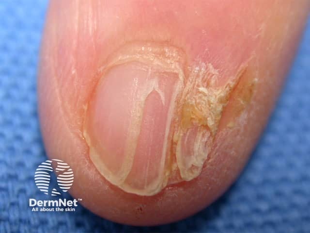
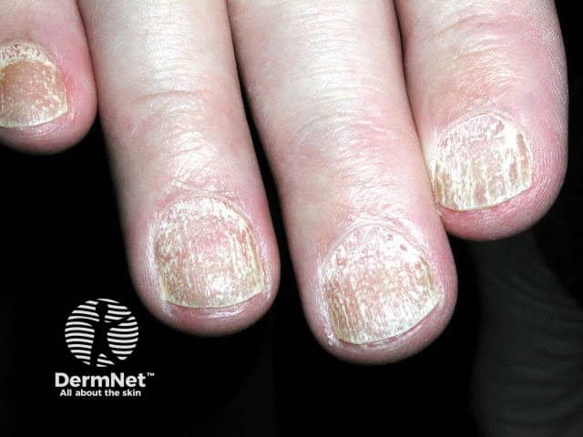
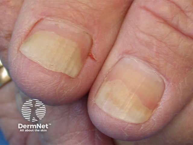
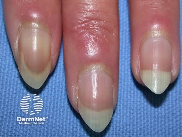
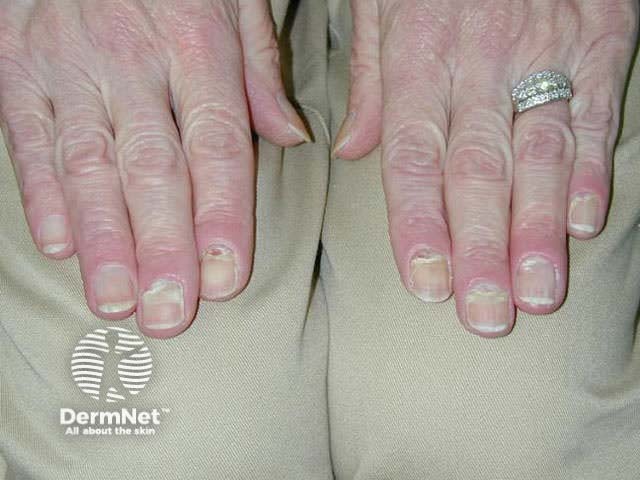
Psoriasis may result in haphazard nail pitting, onycholysis, subungual hyperkeratosis, ridging and/or yellow hypertrophied nail plate.
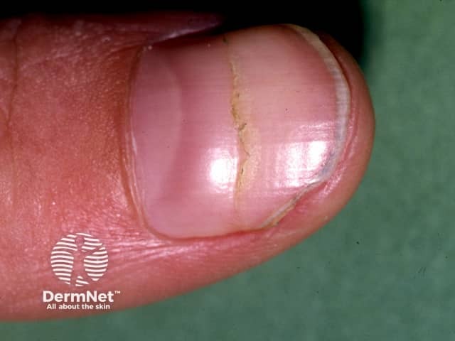
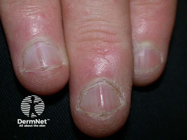
Eczema is associated with irregular pitting, ridging and paronychia.
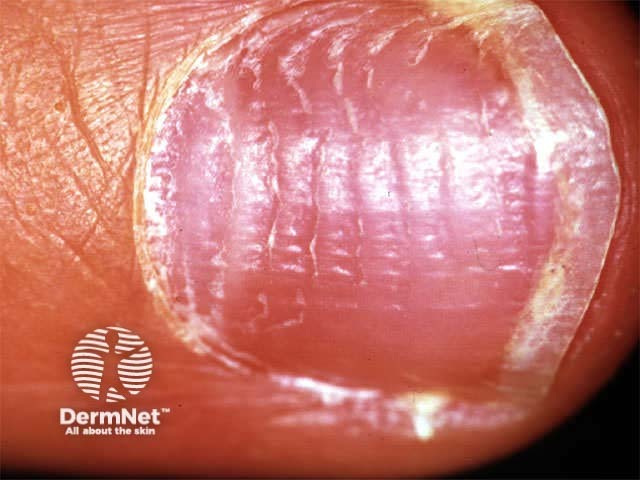
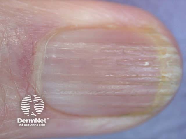
Lichen planus thins the nail plate, which may become grooved and ridged. The nail may darken, thicken or lift off the nail bed.
Nail plate abnormalities are often due to inflammatory conditions affecting the matrix or nail bed. Specific diagnoses may be made from characteristic appearances, which are generally self-explanatory.
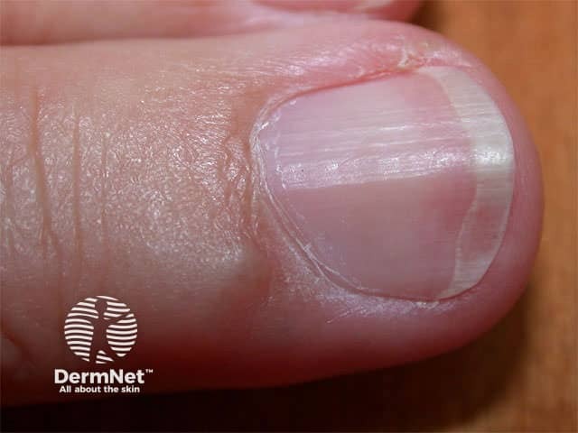
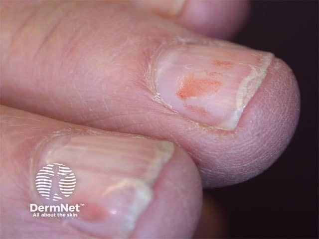
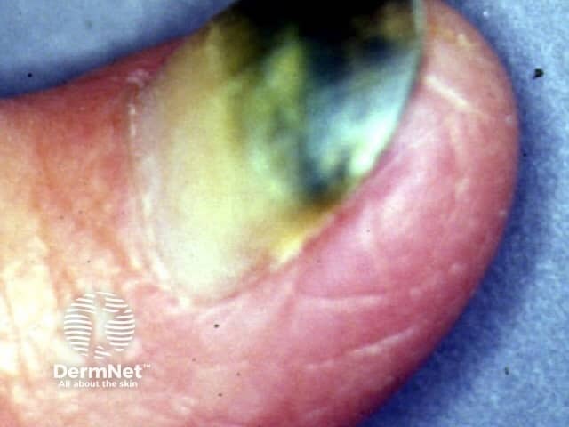
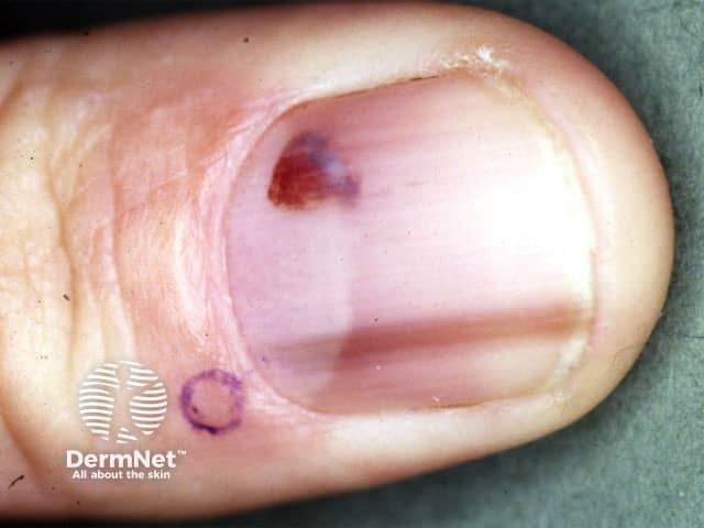
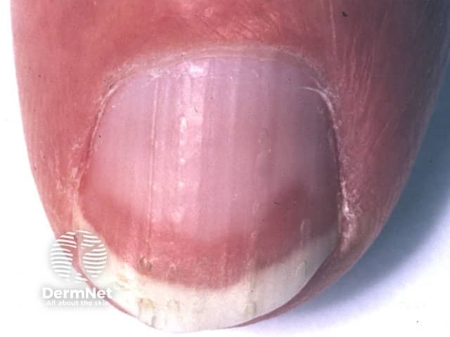
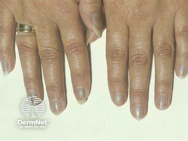
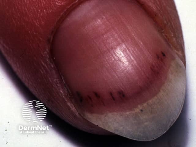
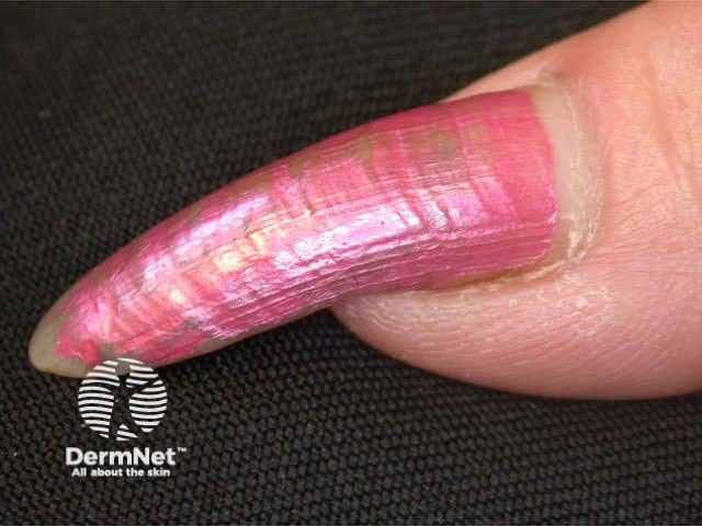
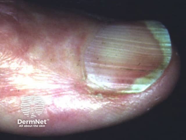
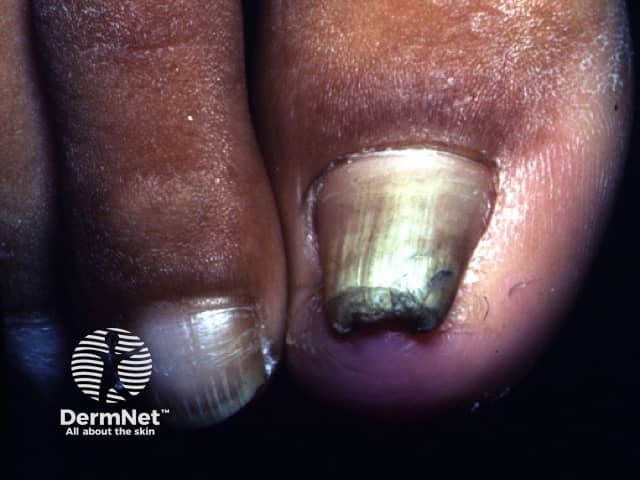
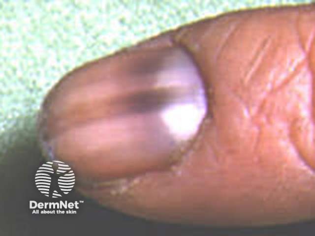
Distinguish a discoloured nail bed from a discoloured nail plate. The most important diagnosis to exclude is subungual melanoma, which presents as a pigmented linear band in the nail plate, which slowly expands at the proximal border and may extend to involve the proximal or lateral nail fold or eponychium.
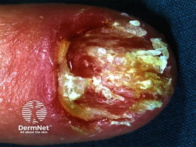
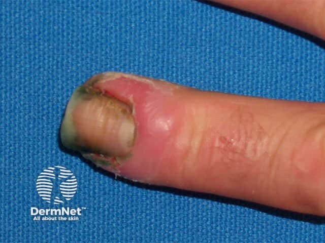
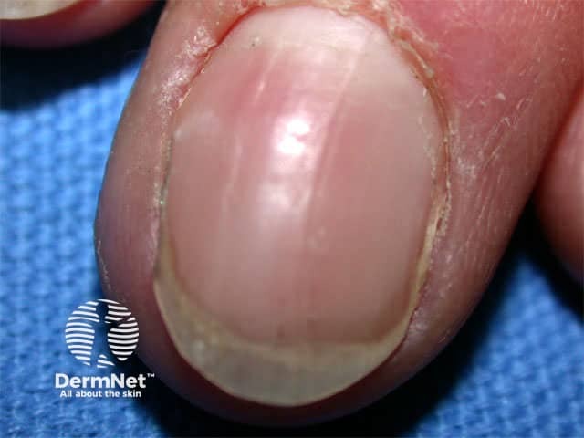
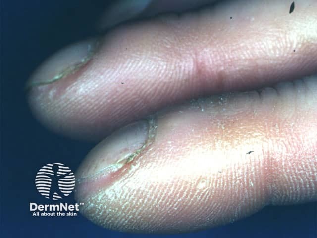
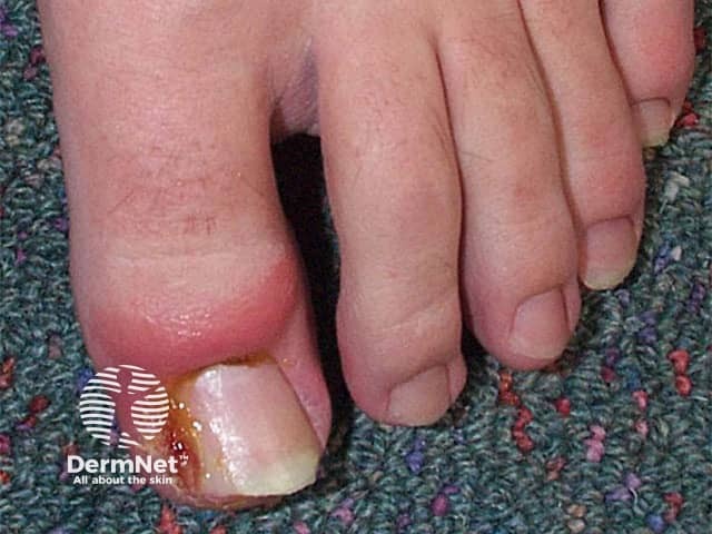
The cuticle (eponychium) is an area of keratin joining the skin of the posterior nail fold to the nail plate. Loss of cuticle results in paronychia: an acute or chronic inflammatory reaction involving nail fold (swelling, tenderness, sometimes pus).
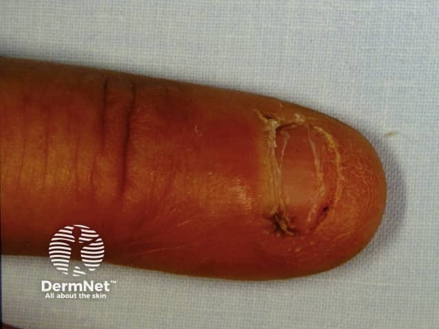
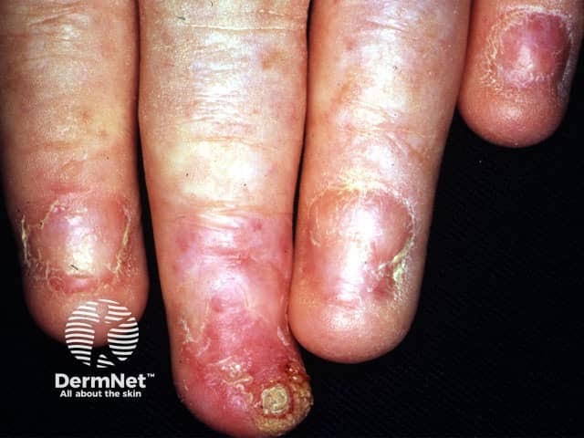
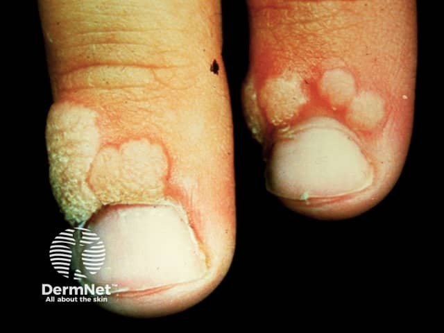
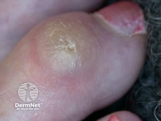
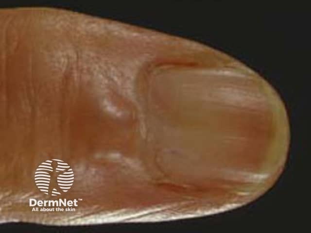
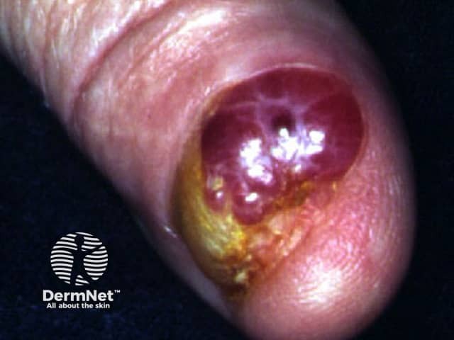
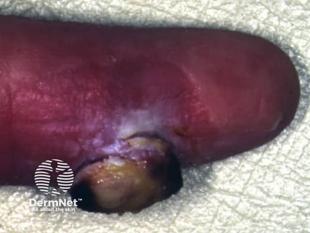
Common skin lesions may arise on the skin close to nails. Benign lesions such as myxoid cyst in the nail matrix area can affect nail growth thus causing a linear depression in the nail plate. Malignant tumours such as squamous cell carcinoma or melanoma can destroy the nail plate.
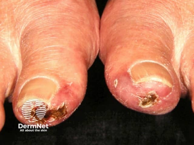
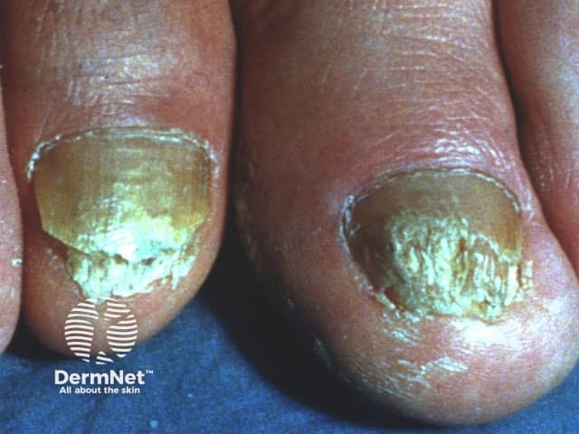
Describe the clinical signs of onychomycosis.
Information for patients

