

Clinical images
Created 2017.
- Practice taking photographs of different body parts and lesions.
- Find or create suitable plain grey, green or blue background to subject.
- Remove extraneous objects.
- Lens should be lined up with the subject.
- Ensure focus + exposure is correct.
- Flash is preferred.
- If too close, back off and zoom.
For each skin lesion:
- Location image or anatomic diagram or both
- Macroscopic (macro) image
- Distance should be 20 cm from the lesion.
- May be useful to have a second image from another angle.
- Repeat image with rule if no anatomic reference structure (eg eye).
- Dermatoscopic (micro) images
- Referrals for pigmented lesions MUST include these.
- Clean and polish lens beforehand, between images and after use .
- Apply fluid to lesion, eg methylated spirits in spray bottle, sanitiser solution/gel (essential for unpolarised, desirable for polarised).
- Contact views are best and should be used whenever possible.
- Polarised and unpolarised views, if available.
- Patient identification image
- You can photograph the consent form.
- Some people photograph an ID label.
- Some people stick an ID label onto the patient besides the lesion; labels with date and rule are available to purchase.
- Delasco.com sells Photo Measuring Labels, and Dermatoscopy stamps, stickers and chart record forms.
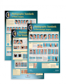
- Poster supplied to DermNet by CanfieldScientific.(PDF file)
- Some smartphone apps have NHI barcode recognition software.
When unable to apply lens in contact with lesion (eg inner canthus):
-
Use non-contact polarised images
Curved surfaces, small body parts are hard to focus and obtain correct exposure:
-
Use grey or green card with cut-out
References
- Finnane A, Curiel-Lewandrowski C, Wimberley G, Caffery L, Katragadda C, Halpern A, Marghoob AA, Malvehy J, Kittler H, Hofmann-Wellenhof R, Abraham I, Soyer HP, On behalf of the International Society of Digital Imaging of the Skin (ISDIS) for the International Skin Imaging Collaboration (ISIC). Proposed Technical Guidelines for the Acquisition of Clinical Images of Skin-Related Conditions. JAMA Dermatol. 2017;153(5):453-457. doi:10.1001/jamadermatol.2016.6214. Journal.
Other websites and resources
- Download: NZ Doctor article 'Clinical imaging: The light and dark arts of skin photography' - PDF file
- Download: NZ Doctor article 'Digital dermatoscopy is a handy alternative in melanoma referrals' - PDF file
- Download: Clinical imaging with an iPhone presentation - PDF file
- New Zealand Teledermatology
- Australian Medical Association “Clinical images and the use of personal mobile devices”
- Waikato DHB for health professionals
On DermNet
ADVERTISEMENT