Main menu
Common skin conditions

NEWS
Join DermNet PRO
Read more
Quick links

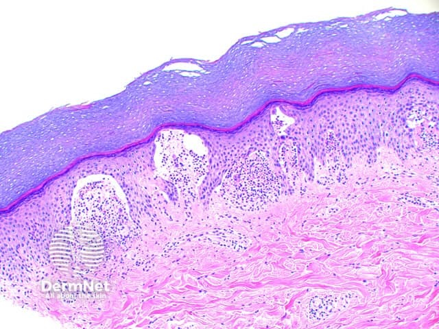
Figure 3
Keywords: Dermatitis herpetiformis, Histopathology-image, Pathology
Scanning power view of dermatitis herpetiformis shows vesicular reaction pattern (Figure 1), characterized by foci or small zones of subepidermal separation (Figures 2 and 3). Dense clusters of neutrophils and scattered eosinophils fill the papillary dermis forming microabscesses (Figure 4). Acantholytic keratinocytes may also be evident within the papillary microabscess (Figure 4). With time the small areas of papillary dermal separation may join to form larger areas of vesiculation.
© DermNet
You can use or share this image if you comply with our image licence. Please provide a link back to this page.
For a high resolution, unwatermarked copy contact us here. Fees apply.
Source: dermnetnz.org