Figure 1
Keywords: Giant cell fibroblastoma, Histopathology-image, Pathology
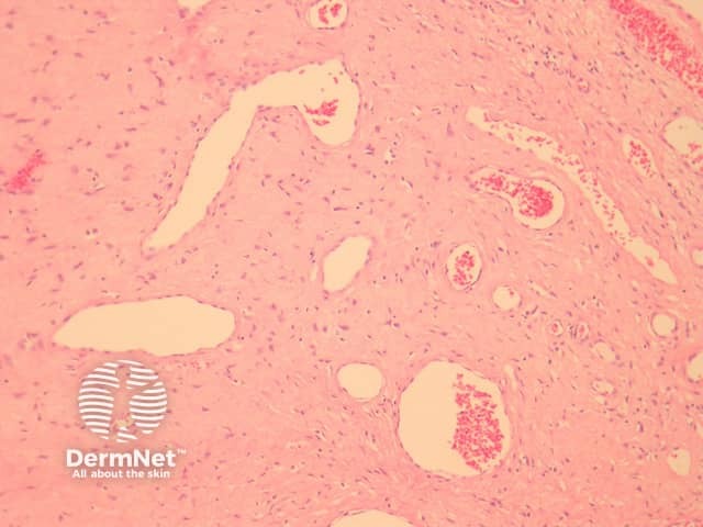
In giant cell fibroblastoma, the tumour is based in the dermis or subcutis. There are striking sinusoidal spaces mimicking blood vessels coursing through the tumour which is set in a myxoid, loose or sclerotic stroma (figure 1). Closer examination reveals the tumour is composed of epithelioid, stellate and spindled cells with occasional multinucleated forms (figures 2, 3). These cells often line the ecstatic vascular spaces. Mitoses are usually rare.
© DermNet
You can use or share this image if you comply with our image licence. Please provide a link back to this page.
For a high resolution, unwatermarked copy contact us here. Fees apply.
Source: dermnetnz.org

