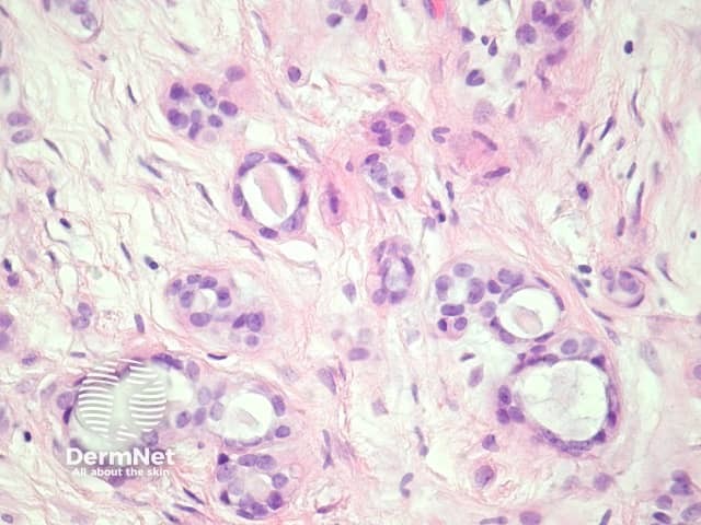
Figure 3
Keywords: Eccrine mixed tumour, Histopathology-image, Pathology
In eccrine mixed tumour, sections show small non-branching ducts set in a fibromucinous or chondroid matrix (figures 1, 2). The tumour is well circumscribed (figure 2) and based in the dermis or subcutis. The ducts are lined by bland cells which at high power resemble syringoma (figure 3). Unusual described features include cribriform glands, osseus metaplasia, and clear cell changes.
© DermNet
You can use or share this image if you comply with our image licence. Please provide a link back to this page.
For a high resolution, unwatermarked copy contact us here. Fees apply.
Source: dermnetnz.org

