Figure 2
Keywords: Histopathology-image
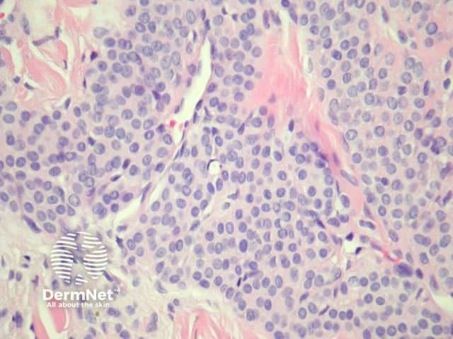
Glomus tumours are dermal, well circumscribed and consist of small vessels surrounded by glomus cells (Figure 1). Glomus cells have bland round to oval nuclei, pale eosinophilic cytoplasm and clearly defined cell margins (Figure 2, 3, 4). Mitotic figures and pleomorphism should not be prominent.
© DermNet
You can use or share this image if you comply with our image licence. Please provide a link back to this page.
For a high resolution, unwatermarked copy contact us here. Fees apply.
Source: dermnetnz.org

