Myofibroma pathology
Keywords: Myofibroma, Histopathology-image, Pathology
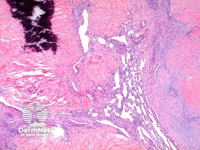
Low power view of myofibroma demonstrates a well defined multinodular tumour arising in the deep dermis or subcutis (Figure 1). Also at low power a branching ‘staghorn’ like pattern of blood vessels can be seen between the tumour nodules (Figure 2). Areas of calcification can often be seen (Figures 2, 3 and 5). The tumour nodules are comprised of a spindle cell proliferation with short plump nuclei (Figures 4,5 and 6). A basophilic tinge in the spindled peripheral component of the nodules is evident (Figures 7 and 8). Sclerotic collagen in the centre of the tumour nodules gives a biphasic appearance to the tumour (Figure 9).
© DermNet
You can use or share this image if you comply with our image licence. Please provide a link back to this page.
For a high resolution, unwatermarked copy contact us here. Fees apply.
Source: dermnetnz.org

