Main menu
Common skin conditions

NEWS
Join DermNet PRO
Read more
Quick links

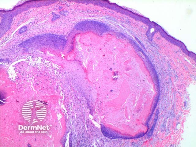
Figure 2
Keywords: Pilomatricoma, Histopathology-image, Pathology
At low power the histological pattern seen in pilomatricoma is of a well-circumscribed nodulocystic tumour (Figure 2). While predominantly seen within the lower dermis, extension into the subcutaneous tissue is not uncommon. The tumour is comprised of a basaloid proliferation resembling the hair matrix cells, which matures into structureless eosinophilic cells lacking nuclei called shadow cells (Figures 3 and 4). The shadow cell area represents differentiation towards the hair cortex. Frequently there are areas of calcification within the shadow cell regions (Figure 5). A histiocytic infiltrate with multinucleated cells forms at sites of rupture (Figure 6).
© DermNet
You can use or share this image if you comply with our image licence. Please provide a link back to this page.
For a high resolution, unwatermarked copy contact us here. Fees apply.
Source: dermnetnz.org