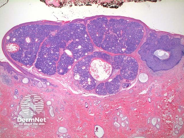
Figure 1
Keywords: Sebaceoma, Histopathology-image, Pathology
Scanning power of the histology of sebaceoma demonstrates a relatively well-circumscribed tumour nodule typically within the deep dermis frequently with attachment to the epidermis. Low power identifies a lobulated tumour, which may demonstrate areas of cyst formation (Figure 1). The tumour is comprised of basaloid cells and a minority of sebaceous cells in addition to small ducts with the crenelated eosinophilic lining seen in sebaceous ducts (Figures 2, 3 and 4). A variable number and distribution of sebocytes is seen, but should remain the minor proportion compared to the basaloid component. Frequent mitoses can be seen in this tumour, though cytological atypia is lacking (Figure 5).
© DermNet
You can use or share this image if you comply with our image licence. Please provide a link back to this page.
For a high resolution, unwatermarked copy contact us here. Fees apply.
Source: dermnetnz.org

