Figure 3
Keywords: Squamous cell carcinoma in situ, Histopathology-image, Pathology
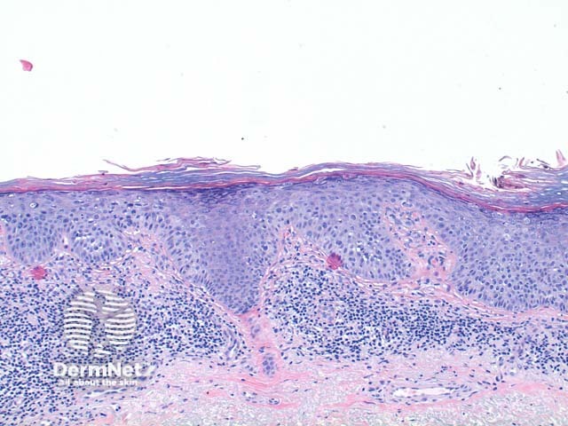
The scanning power view of squamous cell carcinoma in situ (SCCIS) reveals epidermal alteration (Figure 1). Closer inspection reveals atypia of the keratinocytes across the full thickness of the epidermis (Figures 2 and 3). There is a loss of the granular layer and overlying zones of parakeratosis. Sparing of the adnexal ostial epithelium is commonly seen (Figure 3). The keratinocytes show cytologic atypia with disorderly maturation.
© DermNet
You can use or share this image if you comply with our image licence. Please provide a link back to this page.
For a high resolution, unwatermarked copy contact us here. Fees apply.
Source: dermnetnz.org

