Main menu
Common skin conditions

NEWS
Join DermNet PRO
Read more
Quick links

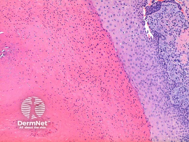
Figure 3
Keywords: Trichilemmal cyst, Histopathology-image, Pathology
Scanning power view of trichilemmal cyst shows a epithelial lined cyst filled with brightly eosinophilic keratinaceous debris (Figure 1). Focal rupture of the cyst may occur with an associated giant cell reaction (Figure 2). Closer inspection of the cyst wall identifies trichilemmal differentiation (Figures 3 and 4) as occurs in the outer root sheath of the hair follicle. This is seen as maturation of squamous epithelium with lack of a granular layer. The eosinophilic keratin centrally is densely packed frequently displaying cholesterol clefts. Focal calcification is seen in around 25% of cases (Figure 5).
© DermNet
You can use or share this image if you comply with our image licence. Please provide a link back to this page.
For a high resolution, unwatermarked copy contact us here. Fees apply.
Source: dermnetnz.org