Main menu
Common skin conditions

NEWS
Join DermNet PRO
Read more
Quick links

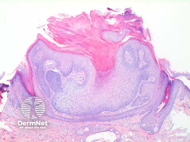
Figure 2
Keywords: Histopathology-image
Trichilemmoma is usually a symmetrical epithelial nodular proliferation. Figure 1. There may be mild papillomatosis with overlying hyperkeratosis. Figure 2. The key finding is of a downgrowth of epithelial cells with increasing clear cell differentiation. Figure 2, Figure 3. These changes are frequently more obvious towards the base of the lesion. The clear cell is PAS positive but diastase labile indicative of the glycogen contents. There is often basal peripheral palisading, resting on a distinctive PASD positive eosinophilic hyaline basement membrane. Figure 4.
© DermNet
You can use or share this image if you comply with our image licence. Please provide a link back to this page.
For a high resolution, unwatermarked copy contact us here. Fees apply.
Source: dermnetnz.org