Figure 3
Keywords: Rheumatoid nodule, Histopathology-image, Pathology
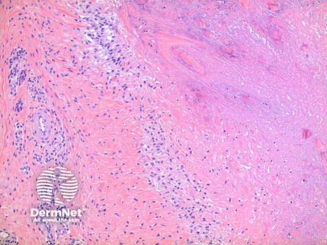
In a rheumatoid nodule, scanning power view reveals a granulomatous tissue reaction pattern (Figure 1). Well formed necrobiotic granulomas form within the dermis frequently with deep extension (Figure 2). There is a surrounding palisade of histiocytes and a mixed infiltrate of lymphocytes, plasma cells, multinucleated giant cells and occasional eosinophils (Figure 3).
© DermNet
You can use or share this image if you comply with our image licence. Please provide a link back to this page.
For a high resolution, unwatermarked copy contact us here. Fees apply.
Source: dermnetnz.org

