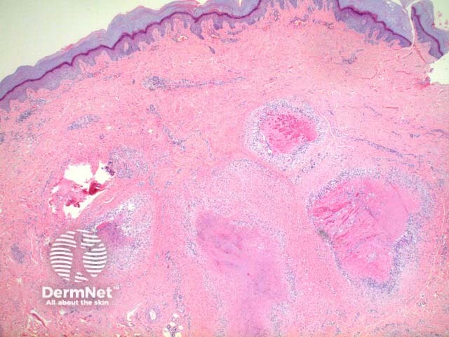
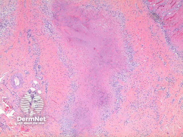
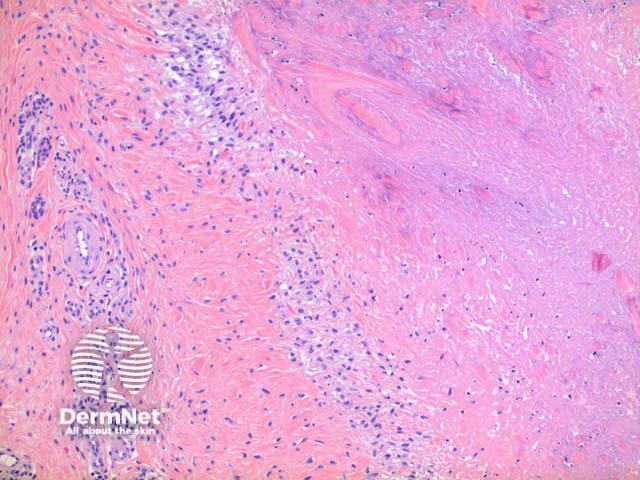
In a rheumatoid nodule, scanning power view reveals a granulomatous tissue reaction pattern (Figure 1). Well formed necrobiotic granulomas form within the dermis frequently with deep extension (Figure 2). There is a surrounding palisade of histiocytes and a mixed infiltrate of lymphocytes, plasma cells, multinucleated giant cells and occasional eosinophils (Figure 3).
Additional staining for fungal infections and mycobacteria should be considered in all significant granulomatous infiltrates.
Deep granuloma annulare: The necrobiotic centres in rheumatoid nodules tend to demonstrate a bright eosinophilic consistency, whereas in granuloma annulare mucin deposition may be seen imparting a basophilic tinge.

