Figure 4
Keywords: Wells syndrome, Histopathology-image, Pathology
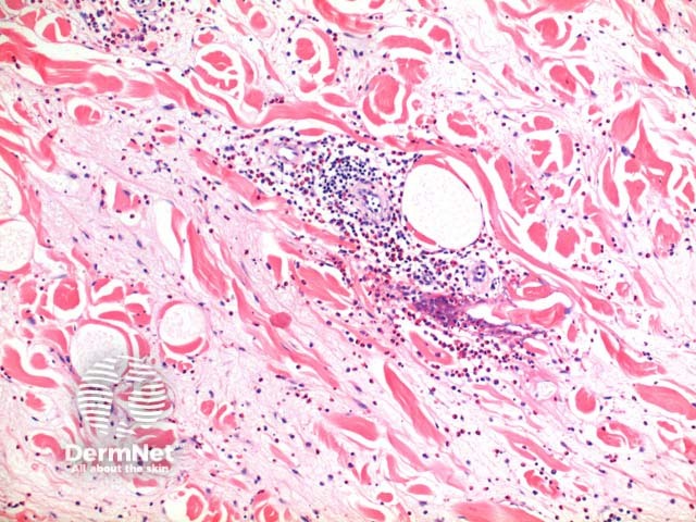
Scanning power view of Wells syndrome reveals a superficial and deep perivascular and interstitial inflammatory pattern (Figures 1 and 2). This can be seen to extend into the subcutaneous tissue (Figure 3) or even the underlying muscle. The inflammatory infiltrate is comprised of lymphocytes, histiocytes and abundant eosinophils (Figures 4,5 and 6). Degranulation of the eosinophils is seen forming flame figures (Figures 4 and 5). In this particular case extensive interstitial mucin is seen (Figures 4 and 5).
© DermNet
You can use or share this image if you comply with our image licence. Please provide a link back to this page.
For a high resolution, unwatermarked copy contact us here. Fees apply.
Source: dermnetnz.org

