Main menu
Common skin conditions

NEWS
Join DermNet PRO
Read more
Quick links

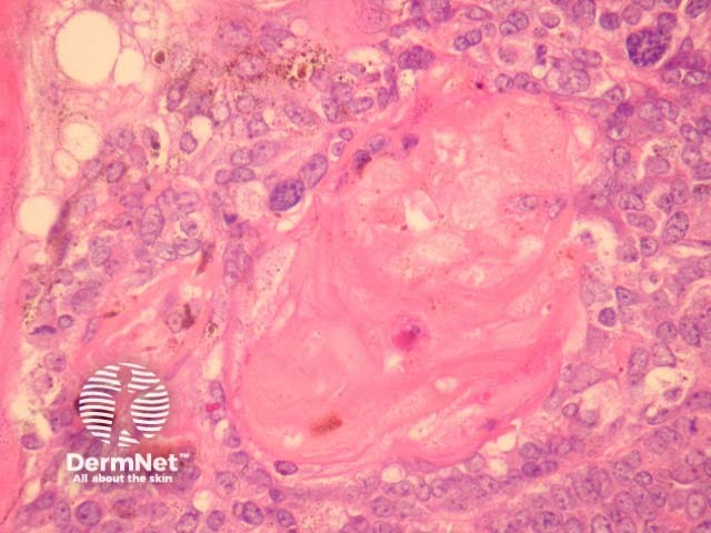
Figure 6
Keywords: Melanocytic matricoma, Histopathology-image, Pathology
Sections through melanocytic matricoma show a well-circumscribed dermal tumour which may encroach and erode the overlying epidermis (figure 1). There is heavy melanin deposition. The tumour is made up of basaloid cells with some nuclear pleomorphism and conspicuous mitotic activity. Intermixed with these epithelial cells are a population of dendritic melanocytes (figures 1-4). There is focal “ghost cell” keratinisation (figures 5, 6).
© DermNet
You can use or share this image if you comply with our image licence. Please provide a link back to this page.
For a high resolution, unwatermarked copy contact us here. Fees apply.
Source: dermnetnz.org